Monday, September 28, 2020

Vessels of the orbit

 Ophthalmic Artery

Origin

The ophthalmic artery is a branch of the cerebral part of the internal carotid artery, given off medial to the anterior clinoid process close to the optic canal

Course and Relations

1. The artery enters the orbit through the optic canal, lying inferolateral to the optic nerve. 

Both the artery and nerve lie in a common dural sheath.

2. In the orbit, the artery pierces the dura mater, ascends over the lateral side of the optic nerve, and 

crosses above the nerve from lateral to medial side along with the nasociliary nerve. 

It then runs forwards along the medial wall of the orbit between the superior oblique and the medial rectus muscles, and parallel to the nasociliary nerve.

3. It terminates near the medial angle of the eye by dividing into the supratrochlear and dorsal nasal branches


Branches

While still within the dural sheath, the ophthalmic artery gives off the central artery of the retina. 

After piercing the dura mater, it gives off a large lacrimal branch that runs along the lateral wall of the orbit. 

The main artery runs towards the medial wall of the orbit giving off a number of branches.


The central artery of retina is the first and most important branch of the ophthalmic artery. 

It first lies below the optic nerve. 

It pierces the dural sheath of the nerve and runs forwards for a short distance between these two. 

It then enters the substance of the nerve and runs forwards in its centre to reach the optic disc 

Here it divides into branches that supply the retina

The central artery of the retina is an end artery.

It does not have effective anastomoses with other arteries.

Occlusion of the artery results in blindness. 

The intraocular part of the artery can be seen, in the living, through an ophthalmoscope.


Branches from Lacrimal artery
1. Branches are given to the lacrimal gland.
2. Two zygomatic branches enter canals in the zygomatic bone. One branch appears on the face
through the zygomaticofacial foramen. The other appears on the temporal surface of the bone 
through the zygomaticotemporal foramen.
3. Lateral palpebral branches supply the eyelids.
4 A recurrent meningeal branch runs backwards to enter the middle cranial fossa through the superior orbital fissure.
5. Muscular branches supply the muscles of the orbit.


1. The posterior (long and short) ciliary arteries supply chiefly the choroid and iris. 
The eyeball is also supplied through anterior ciliary branches which are given off from arteries supplying 
muscles attached to the eyeball.
2. The supraorbital and supratrochlear branches supply the skin of the forehead.
3. The anterior and posterior ethmoidal branches enter foramina in the medial wall of the orbit to supply the ethmoidal air sinuses. 
They then enter the anterior cranial fossa. 
The terminal branches of the anterior artery enter the nose and supply part of it.
4. The medial palpebral branches supply the eyelids.
5. The dorsal nasal branch supplies the upper part of the nose.


Clinical Anatomy
The anterior ciliary arteries arise from the muscular branches of ophthalmic artery. 
The muscular arteries are important in this respect.
The central artery of retina is the only arterial supply to most of the nervous layer, the retina of the eye. If this artery is blocked, there is sudden blindness.


Ophthalmic Veins
The superior ophthalmic vein: 
  • It accompanies the ophthalmic artery. 
  • It lies above the optic nerve. 
  • It receives tributaries corresponding to the branches of the artery, 
  • passes through the superior orbital fissure, and drains into the cavernous sinus. 
  • It communicates anteriorly with the supraorbital and angular veins


The Inferior ophthalmic vein: 
It runs below the optic nerve.
It receives tributaries from the lacrimal sac, the lower orbital muscles, and the eyelids, and 
ends either by joining the superior ophthalmic vein or drains directly into the cavernous sinus. 
It communicates with the pterygoid plexus of veins by small veins passing through the inferior orbital fissure.


Watch the lectures on YouTube:
Blood vessels of the Orbit | Ophthalmic artery & branches | Superior & Inferior Ophthalmic Veins


Friday, September 25, 2020

Extraocular muscles

 TYPES OF EXTRAOCULAR MUSCLES

Voluntary Muscles

1. Four recti:

a. Superior rectus.

b. Inferior rectus.

c. Medial rectus.

d. Lateral rectus.

2. Two obliqui:

a. Superior oblique.

b. Inferior oblique.

3 The levator palpebrae superioris elevates the upper eyelid.


lnvoluntary Muscles

1. The superior tarsal muscle is the deeper portion of the levator palpebrae superioris. 

It is inserted on the upper margin of the superior tarsus. 

It elevates the upper eyelid.

2. The inferior tarsal muscle extends from the fascial sheath of the inferior rectus and inferior 

oblique to the lower margin of the inferior tarsus. 

It possibly depresses the lower eyelid.

3. The orbitalis bridges the inferior orbital fissure. 

Its action is uncertain


Voluntary Muscles
Origin
1. The four recti arise from a common annular tendon of tendinous ring of zinn. 
The ring is attached to the middle part of superior orbital fissure.
The lateral rectus has an additional small tendinous head which arises from the orbital surface of the greater wing of the sphenoid bone lateral to the tendinous ring. 
Through the gap between the two heads abducent nerve 
passes.
2. The superior oblique arises from the undersurface of lesser wing of the sphenoid, superomedial to the optic canal.
3. The inferior oblique arises from the orbital surface of the maxilla, lateral to the lacrimal groove. The muscle is situated near the anterior margin of the orbit.
4. The levator palpebrae superioris arises from the orbital surface of the lesser wing of the sphenoid bone, 
anterosuperior to the optic canal and to the origin of the superior rectus.


Insertion
1.The recti are inserted into the sclera, a little posterior to the limbus (corneo-scleral junction). 
The average distances of the insertions from the cornea are:
superior 7.7 mm; 
inferior 6.5 mm, 
medial 5.5 mm;
lateral 6.9 mm.
2. The tendon of the superior oblique passes through a fibrocartilaginous pulley attached to the trochlear fossa of the frontal bone. 
The tendon then passes laterally, downwards and backward below the superior rectus. 
It is inserted into the sclera behind the equator of the eyeball, between the superior rectus and the lateral rectus.
3. The inferior oblique is fleshy throughout. 
It passes laterally, upwards and backwards below the inferior rectus and then deep to the lateral rectus. 
The inferior oblique is inserted close to the superior oblique a little below and posterior to the latter.
4. The flat tendon of the levator splits into a superior or voluntary and an inferior or involuntary lamellae.
The superior lamella of the levator is inserted into the anterior surface of the superior tarsus, and into the skin of the upper eyelid. 
The inferior lamella (smooth part) is inserted into the upper margin of the superior tarsus and into superior
conjunctival fornix.


Nerve Supply
1. The superior oblique is supplied by the IV cranial or trochlear nerve (SO4).
2. The lateral rectus is supplied by the VI cranial or abducent nerve (LR6).
3. The remaining five extraocular muscles; superior, inferior and medial recti; inferior oblique and part
of levator palpebrae superioris are all supplied by the III cranial or oculomotor nerve


Actions
1. The movements of the eyeball are as follows.
a. Around a transverse axis . (Y- axis)
Upward rotation or elevation (33').
Downwards rotation or depression (33’).
b. Around a vertical axis . (X- axis)
Medial rotation or adduction (50").
Lateral rotation or abduction (50').
c. Around an anteroposterior axis (Z- axis)
Intortion
Extortion.
The rotatory movements of the eyeball upwards, downwards, medially or laterally, are defined in terms of 
the direction of movement of the centre of the pupil. 
The tortions are defined in terms of the direction of movement of the upper margin of the pupil at 12 o'clock position.
d. The movements given above can take place in various combinations.


Single or pure movements are produced by combined actions of muscles. 
Similar actions get added together, while opposing actions cancel each other enabling pure movements.
a. Upward rotation or elevation: 
By the superior rectus and the inferior oblique.
b. Downward rotation or depression: 
By the inferior rectus and the superior oblique.
c. Medial rotation or adduction; 
By the medial rectus, the superior rectus and the inferior rectus.
d. Lateral rotation or abduction: 
By the lateral rectus, the superior oblique and the inferior oblique.
e. Intortion: By the superior oblique and the superior rectus.
f. Extortion: By the inferior oblique and the inferior rectus.


Combined movements of the eyes
Normally, movements of the two eyes are harmoniously coordinated. 
Such coordinated movements of both eyes are called conjugate ocular movements


Clinical Anatomy
1. Weakness or paralysis of a muscle causes squint or strabismus, 
which maybe concomitant or paralytic.
Concomitant squint is congenital; 
there is no limitation of movement, and no diplopia
In paralytic squint, movements are limited,
diplopia and vertigo are present, head is turned in the direction of the function of paralysed muscle, and there is a false orientation of the field of vision.
Nystagmus is characterized by involuntary, rhythmical oscillatory movements of the eyes. 
This is due to incoordination of the ocular muscles. 
It may be either vestibular or cerebellar/ or even congenital.


Watch lectures on YouTube:


Thursday, September 24, 2020

Orbits & its contents

 Features

  • The orbits are pyramidal cavities, 
  • situated one on each side of the root of the nose. 
  • They provide sockets for rotatory movements of the eyeball. 
  • The long axis of the each orbit passes backwards and medially. 
  • The medial walls are parallel to each other at a distance of 2.5 cm
  • but the lateral walls are set at right angles to each other.


Contents

1. Eyeball: Eyeball occupies anterior one-third of orbit.
2. Fascia: Orbital and bulbar.
3. Muscles: Extraocular and intraocular.
4. Vessels: Ophthalmic artery, superior and inferior ophthalmic veins, and lymphatics.
5. Nerves: Optic, oculomotor, trochlear and abducent; branches of ophthalmic and maxillary nerves, and sympathetic nerves.
6. Lacrimal gland: 
7. Orbital fat.

Visual Axis ond Orbital Axis
Axis passing through centres of anterior and posterior poles of the eyeball is known as visual axis. 
It makes an angle of 20 - 25˚ with the orbital axis, (line passing through optic canal and centre of base of orbit,)


Orbital Fascia or Periorbita
  • It forms the periosteum of the bony orbit. 
  • Due to the loose connection to bone, it can be easily stripped.
  • Posteriorly, it is continuous with the dura mater and with the sheath of the optic nerve. 
  • Anteriorly, it is continuous with the periosteum lining the bones around the orbital margin.
  • There is a gap in the periorbita over the inferior orbital fissure. 
  • This gap is bridged by connective tissue with some smooth muscle fibres in it. 
  • These fibres constitute the orbitalis muscle.
  • a. At the upper and lower margins of the orbit, the orbital fascia sends off flap-like continuations into the eyelids. These extensions form the orbital septum.
  • b. A process of the fascia holds the fibrous pulley of the tendon of the superior oblique muscle in place.
  • c. Another process forms the lacrimal fascia which bridges the lacrimal groove.


Fascial Sheath of Eyeball of Bulbar Fascia
1. Tenon's capsule forms a thin, loose membranous sheath around the eyeball, extending from the optic nerve to the sclerocorneal junction or limbus. 
It is separated from the sclera by the episcleral space which is traversed by delicate fibrous bands. 
The eyeball can freely move within this sheath.
2. The sheath is pierced by:
a. Tendons of the various extraocular muscles.
b. Ciliary vessels and nerves around the entrance of the optic nerve.
3. The sheath gives off a number of expansions.
a. A tubular sheath covers each orbital muscle.
b. The medial check ligament is a strong triangular expansion from the sheath of the medial rectus muscle; it is attached to the lacrimal bone.
c. The lateral check ligament is a strong triangular expansion from the sheath of the lateral rectus muscle; it is attached to the zygomatic bone
4. The lower part of Tenon's capsule is thickened, and is named the suspensory ligament of the eye or the
suspensory ligament of Lockwood .
It is expanded in the centre and narrow at its extremities, and is slung like a hammock below the eyeball. 
It is formed by union-of the margins of the sheaths of the inferior rectus and the inferior oblique muscles with the medial and lateral check ligaments.


Watch lectures on YouTube:


Tuesday, September 22, 2020

Petrosal Nerves

 1. The greater petrosal nerve 

  • carries gustatory and parasympathetic fibres. 
  • It arises from the geniculate ganglion of the facial nerve, and 
  • enters the middle cranial fossa through the hiatus for the greater petrosal nerve on the anterior surface of the petrous temporal bone. 
  • It proceeds towards the foramen lacerum,  where it joins the deep petrosal nerve which carries sympathetic fibres to form the nerve of the pterygoid canal
  • The nerve of the pterygoid canal passes through the pterygoid canal to reach the pterygopalatine
  • ganglion. 
  • The parasympathetic fibres relay in this ganglion. 
  • Postganglionic parasympathetic fibres arising in the ganglion ultimately supply the lacrimal gland 
  • and the mucosal glands of the nose, palate and pharynx.
  • The gustatory or taste fibres do not relay in the ganglion and are distributed to the palate.


2. The deep petrosal nerve
  • sympathetic in nature, 
  • is a branch of the sympathetic plexus around the internal carotid artery. 
  • It contains postganglionic fibres from the superior cervical sympathetic ganglion. 
  • The nerve joins the greater petrosal nerve to form the nerve of the pterygoid canal. 
  • The sympathetic fibres in it are distributed through the branches of the pterygopalatine ganglion


3. The lesser petrosal nerve, 
  • parasympathetic in nature,
  • is a branch of the tympanic plexus, deriving its preganglionic parasympathetic fibres from the tympanic branch of the glossopharyngeal nerve. 
  • It emerges through the hiatus for the lesser petrosal nerve, 
  • situated just lateral to the hiatus for the greater petrosal nerve, 
  • passes out of the skull through the foramen ovale, and ends in the otic ganglion 
  • Postganglionic fibres arising in the ganglion supply the parotid gland through the auriculotemporal nerve


4. The external petrosal nerve, 
  • sympathetic in nature is an inconstant branch from the sympathetic plexus around the middle meningeal artery to the geniculate ganglion of the facial nerve.


Friday, September 18, 2020

Middle Meningeal Artery

The middle meningeal artery is important to the surgeon because this artery is the commonest source of 

extradural haemorrhage, which is an acute surgical emergency

The artery is a branch of the first part of the maxillary artery, given off in the infratemporal fossa 

Course ond Relations

1. In the infratemporal fossa, the artery runs upwards and medially deep to the lateral pterygoid muscle and superficial to the sphenomandibular ligament.

Here it passes through a loop formed by the two roots of the auriculotemporal nerve.

2. It enters the middle cranial fossa through the foramen spinosum


3. In the middle cranial fossa, the artery has an extradural course, but the middle meningeal veins are closer to the bone than the artery. 
Here the artery runs forwards and laterally for a variable distance, grooving the squamous temporal bone, and divides into a frontal and parietal branch 
4. The frontal or anterior branch is larger than the parietal branch. 
First it runs forwards and laterally towards the lateral end of the lesser wing of the sphenoid. Then it runs obliquely upwards and backwards, parallel to, and a little in front of the central sulcus of the cerebral hemisphere. 
Thus after crossing the pterion, the artery is closely related to the motor area of the cerebral cortex.
5. The parietal or posterior branch runs backwards over, or near, the superior temporal sulcus of the cerebrum, about 4 cm above the level of the zygomatic arch. 
It ends in front of the posteroinferior angle of the parietal bone by dividing into branches.


Branches
The middle meningeal artery supplies only small branches to the dura mater. 
It is predominantly a periosteal artery supplying bone and red bone marrow in the diploe.
Within the cranial cavity, it gives off:
a. The ganglionic branches to the trigeminal ganglion.
b. A petrosal branch to the hiatus for the greater petrosal nerve.
c. A superior tympanic branch to the tensor tympani.
d. Temporal branches to the temporal fossa.
e. Anastomotic branch that enters the orbit and anastomoses with the lacrimal artery


Clinical Anatomy

  • The middle meningeal artery is of great surgical importance because it can be torn in head injuries resulting in extradural haemorrhage. 
  • The frontal or anterior branch is commonly involved. 
  • The haematoma presses on the motor area, giving rise to hemiplegia of the opposite side. 
  • The anterior division can be approached surgically by making a hole in the skull over the pterion, 4 cm above the midpoint of the zygomatic arch.
  • Rarely, the parietal or posterior branch is implicated, causing contralateral deafness. In this case, the hole is made at a point 4 cm above and 4 cm behind the external acoustic meatus
Watch lectures on YouTube:


Wednesday, September 16, 2020

Trigeminal Ganglion

 This is the sensory ganglion of the fifth cranial nerve. It is homologous with the dorsal nerve root ganglia of spinal nerves. 

All such ganglia are made up of pseudounipolar nerve cells, with a 'T'-shaped arrangement of their process; one process arises from the cell body which then divides into a central and a peripheral process

The ganglion is crescentic or semilunar in shape, with its convexity directed anterolaterally. 

The three divisions of the trigeminal nerve emerge from this convexity. 

The posterior concavity of the ganglion receives the sensory root of the nerve

Situation and Meningeal Relations

The ganglion lies on the trigeminal impression, on the anterior surface of the petrous temporal bone near its apex. 

It occupies a special space of dura mater, called the trigeminal or Meckel's cave. 

There are two layers of dura below the ganglion. 

The cave is lined by pia-arachnoid, so that the ganglion along with the motor root of the trigeminal nerve is surrounded by CSF. 

The ganglion lies at a depth of about 5 cm from the preauricular point.


Relations

Medially
1. Internal carotid artery.
2. Posterior part of cavernous sinus.

Laterally
1. Middle meningeal artery.

Superiorly
1. Parahippocampal gyrus.

Inferiorly
1. Motor root of trigeminal nerve.
2. Greater petrosal nerye.
3. Apex of the petrous temporal bone.
4. The foramen lacerum

Associated Root and Branches
The central processes of the ganglion cells form the large sensory root of the trigeminal nerve which is attached to pons at its junction with the middle cerebellar peduncle.
The peripheral processes of the ganglion cells form three divisions of the trigeminal nerve, namely the
ophthalmic, maxillary and mandibular.
The small motor root of the trigeminal nerve is attached to the pons superomedial to the sensory root.
It passes under the ganglion from its medial to the lateral side, and joins the mandibular nerve at the
foramen ovale.


Blood Supply
The ganglion is supplied by twigs from:
1. Internal carotid
2. Middle meningeal
3. Accessory meningeal arteries
4. By the meningeal branch of the ascending pharyngeal artery.

Clinical Anatomy
1. Intractable facial pain due to trigeminal neuralgia or carcinomatosis may be abolished by injecting alcohol into the ganglion. Sometimes cutting of the sensory root is necessary.
2. Congenital cutaneous naevi on the face (port wine stains) map out accurately the areas supplied by one or more divisions of the V cranial nerve.


Watch lectures on YouTube:


Tuesday, September 15, 2020

Hypophysis cerebri/ Pituitary Gland

The hypophysis cerebri is a small endocrine gland situated in relation to the base of the brain. 

It is often called the master of the endocrine orchestra because it produces a number of hormones which control the secretions of many other endocrine glands of the body

The gland lies in the hypophyseal fossa or sella turcica or pituitary fossa. 

The fossa is roofed by the diaphragma sellae. 

The stalk of the hypophysis cerebri pierces the diaphragma sellae and is attached above to the floor of the third ventricle.

The gland is oval in shape, and measures 8 mm anteroposteriorly and 72 mm transversely. 

It weighs about 500 mg.


Relations
Superiorly
1. Diaphragma sellae 
2. Optic chiasma.
3. Tubercinerium.
4. Infundibular recess of the third ventricle.

Inferiorly
1. Irregular venous channels between the two layers of dura mater lining the floor of the hypophyseal fossa.
2. Hypophyseal fossa.
3. Sphenoidal air sinuses.

On each side
The cavernous sinus with its contents


Subdivisions / Parts and Development
The gland has two main parts:
 Adenohypophysis and neurohypophysis which differ from each other embryologically, 
morphologically and functionally. 

The adenohypophysis develops as an upward growth called the Rathke's pouch from the ectodermal roof of the stomodeum. 
The neurohypophysis develops as a downward growth from the floor of the diencephalon,
and is connected to the hypothalamus by neural pathways.


Adenohypophysis
1. Anterior lobe or pars anterior, Pars distalis, or pars glandularis: 
This is the largest part of the gland

2. lntermediate lobe or pars intermedia: 
This is in the form of a thin strip which is separated from the anterior lobe by an intraglandular cleft, a remnant of the lumen of Rathke's pouch.

3. Tuberal lobe or pars tuberalis: 
It is an upward extension of the anterior lobe that surrounds and forms part of the infundibulum.


Neurohypophysis
1. Posterior lobe or neural lobe, pars posterior: 
It is smaller than the anterior lobe and lies in the posterior concavity of the larger anterior lobe.
2. lnfundibular stem, which contains the neural connections of the posterior lobe with the hypothalamus.
3. Median eminence of the tubercinerium which is continuous with the infundibular stem


Arterial Supply
The hypophysis cerebri is supplied by the following branches of the internal carotid artery.
1. One superior hypophyseal artery on each side
2. One inferior hypophyseal artery on each side. 
Each superior hypophyseal artery supplies:
a. Ventral part of the hypothalamus.
b. Upper part of the infundibulum.
c. Lower part of the infundibulum through a separate long descending branch, called the trabecular artery.
Each inferior hypophyseal artery divides into medial and lateral branches which join one another to form an arterial ring around the posterior lobe. 
Branches from this ring supply the posterior lobe and also anastomose with branches from the superior hypophyseal artery.
The anterior lobe or pars distalis is supplied exclusively by portal vessels arising from capillary tufts formed by the superior hypophyseal arteries. 
The long portal vessels drain the median eminence and the upper infundibulum, and the short portal vessels drain the lower infundibulum. 
The portal vessels are of great functional importance because they carry the hormone releasing factors from the hypothalamus to the anterior lobe where they control the secretory cycles of different glandular cells.


Venous Drainage
Short veins emerge on the surface of the gland and drain into neighbouring dural venous sinuses. 
The hormones pass out of the gland through the venous blood, and are carried to their target cells.

Hormones
Anterior lobe
Chromophilic cells 50%.
1. Acidophils/alpha-cells; about 43%
a. Somatotrophs: Secrete growth hormone (STH,GH).
b. Mammotrophs (prolactin cells): Secrete lactogenic hormone.
c. Corticotrophs: Secrete ACTH.
2. Basophils/beta-cells, about 7% of cells
a. Thyrotrophs: Secrete TSH.
b. Gonadotrophs: Secrete FSH.
c. Luteotrophs: Secrete LH or ICSH.
Chromophobic cells 50% represent the non-secretory phase of the other cell types, or their precursors.
Intermediate Lobe
It is made up of numerous basophil cells, and chromophobe cells surrounding masses of colloid material. 
It secretes the melanocyte stimulating hormone (MSH).


Posterior Lobe
It is composed of:
1. A large number of nonmyelinated fibres hypothalamo- hypophyseal tract.
2. Modified neurological cells, called pituicytes. They have many dendrites which terminate on or near the
sinusoids.
Hypothalamo-hypophyseal portal system
The hypothalamo-hypophyseal tract begins in the preoptic and paraventricular nuclei of the hypothalamus.
Its short fibres terminate in relation to capillary tufts of portal vessels, providing the possibility for a neural control of the secretory activity of the anterior lobe. 
The long fibres of the neurosecretory tract pass to the posterior lobe and terminate near vascular sinusoids.

The hormones related to the posterior lobe are:
a. Vasopressin (ADH) which acts on kidney tubules.
b. Oxytocin which promotes contraction of the uterine and mammary smooth muscle.
These hormones are actually secreted by the hypothalamus, from where these are transported through the 
hypothalamo-hypophyseal tract to the posterior lobe of the gland.

Clinical Anatomy
Pituitary tumours give rise to two main categories of symptoms:
A. General symptoms due to pressure over surrounding structures:
a. The sella turcica is enlarged in size.
b. Pressure over the central part of optic chiasma causes bitemporal hemianopia
c. Pressure over the hypothalamus may cause one of the hypothalamic syndromes like obesity of Frolich's syndrome in cases with Rathke's pouch tumours.
d. A large tumour may press upon the third ventricle, causing a rise in intracranial pressure.
B. Specific symptoms depending on the cell type of the tumour.
a. Acidophil or eosinophil adenoma causes acromegaly in adults and gigantism in younger patients.
b. Basophil adenoma causes Cushing's syndrome.
c. Chromophobe adenoma causes effects of hypopituitarism.
d. Posterior lobe damage causes diabetes insipidus, although the lesion in these cases usually lies in the hypothalamus.


Watch Lectures on YouTube:
Pituitary gland | Hypophysis Cerebri |Parts |Development |Histology & Function |Blood & Nerve supply


Friday, September 4, 2020

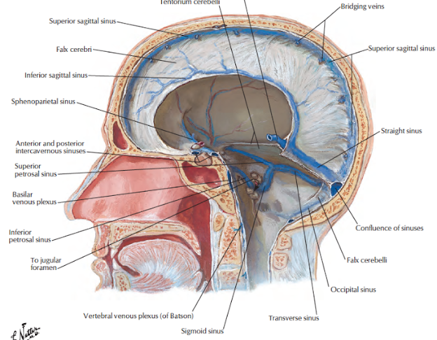
Cranial Venous Sinuses

  • These are venous spaces, the walls of which are formed by dura mater. 
  • They have an inner lining of endothelium. 
  • There is no muscle in their walls.
  • They have no valves.
  • Venous sinuses receive venous blood from the brain, the meninges, and bones of the skull
  • Cerebrospinal fluid is poured into some of them.
  • Cranial venous sinuses communicate with veins outside the skull through emissary veins. 
  • These communications help to keep the pressure of blood in the sinuses constant
  • There are 23 venous sinuses, of which 8 are paired and 7 are unpaired.

Paired Sinuses

There is one sinus each on right and left side.

1. Cavernous sinus.

2. Superior petrosal sinus

3. Inferior petrosal sinus.

4. Transverse sinus

5. Sigmoid sinus.

6. Sphenoparietal sinus.

7. Petrosquamous sinus.

8. Middle meningeal sinus/veins.

Unpaired Sinuses

These are median in position.

1. Superior sagittal sinus.

2. Inferior sagittal sinus.

3. Straight sinus.

4. Occipital sinus.

5. Anterior intercavernous sinus.

6. Posterior intercavernous sinus.

7. Basilar plexus of veins.


Cavernous Sinus

Introduction
  • Each cavernous sinus is a large venous space situated in the middle cranial fossa, 
  • on either side of the body of the sphenoid bone. 
  • Its interior is divided into a number of spaces or caverns by trabeculae. 
  • The trabeculae are much less conspicuous in the living than in the dead.
  • The floor and medial wall of the sinus is formed by the endosteal dura mater. 
  • The lateral wall, and roof are formed by the meningeal dura mater.
  • Anteriorly, the sinus extends up to the medial end of the superior orbital fissure and
  • posteriorly, up to the apex of the petrous temporal bone. 
  • It is about 2 cm long, and 1 cm wide.

Relations
Structures outside the sinus:
1. Superiorly: Optic tract, optic chiasma, olfactory tract, internal carotid artery and anterior perforated
substance.
2. Inferiorly: Foramen lacerum and the junction of the body and greater wing of the sphenoid bone 
3. Medially; Hypophysis cerebri and sphenoidal air sinus.
4. Laterally: Temporal lobe with uncus.
5. Below laterally: Mandibular nerve
6. Anteriorly; Superior orbital fissure and the apex of the orbit.
7. Posteriorly; Apex of the petrous temporal and the crus cerebri of the midbrain.


Structures with in the Lateral Wall 
From above downwards
1. Oculomotor nerve: In the anterior part of the sinus, it divides into superior and inferior divisions which leave the sinus by passing through the superiororbital fissure.
2. Trochlear nerve: In the anterior part of the sinus, it crosses superficial to the oculomotor nerve, and enters the orbit through the superior orbital fissure.
3. Ophthalmic nerve: In the anterior part of the sinus, it divides into the lacrimal, frontal and nasociliary nerves.
4. Maxillary Nerve: It leaves the sinus by passing through the foramen rotundum on its way to the pterygopalatine fossa.
5. Trigeminal ganglion: The ganglion and its dural cave project into the posterior part of the lateral wall of the sinus.

Structures passing through the medial aspect of the sinus:
a. lnternal carotid artery with the venous and sympathetic plexus around it.
b. Abducent nerve, inferolateral to the internal carotid artery.

The structures in the lateral wall and on the medial aspect of the sinus are separated from blood by the endothelial lining.


Tributaries or Incoming Channels
From the orbit
1. The superior ophthalmic vein.
2. A branch of the inferior ophthalmic vein or sometimes the vein itself.
3. The central vein of the retina may drain either into the superior ophthalmic vein or into the cavernous
sinus
From the Brain
1. Superficial middle cerebral vein.
2. Inferior cerebral veins from the temporal lobe
From the Meninges
1. Sphenoparietal sinus.
2. The frontal trunk of the middle meningeal vein may drain either into the pterygoid plexus through the
foramen ovale or into the sphenoparietal or cavernous sinus.


Draining Channels or Communications
The cavernous sinus drains:
1. Into the transverse sinus through the superior petrosal sinus.
2. Into the internal jugular vein through the inferior petrosal sinus and through a plexus around the
internal carotid artery.
3. Into the pterygoid plexus of veins through the emissary veins passing through the foramen ovale,
the foramen lacerum and the emissary sphenoidal foramen.
4. Into the facial vein through the superior ophthalmic vein.
5. The right and left cavernous sinuses communicate with each other through the anterior and posterior
intercavernous sinuses and through the basilar plexus of veins 
All these communications are valveless, and blood can flow through them in either direction.


Factors helping expulsion of blood from the Sinus
1. Expansile pulsations of the internal carotid artery within the sinus.
2. Gravity.
3. Position of the head.

Clinical Anatomy
1. Thrombosis of the cavernous sinus may be caused by sepsis in the dangerous area of the face, in nasal 
cavities, and in paranasal air sinuses. This gives rise to the following symptoms.
a. Nervous symptoms:
- Severe pain in the eye and forehead in the area of distribution of ophthalmic nerve.
- Involvement of the third, fourth and sixth cranial nerves resulting in paralysis of the muscles supplied.
b. Venous symptoms: Marked oedema of eyelids, cornea and root of the nose, with exophthalmos due to 
congestion of the orbital veins.
2. A communication between the cavernous sinus and the internal carotid artery may be produced
by head injury. When this happens the eyeball protrudes and pulsates with each heart beat. It is called the 
pulsating exophthalmos.

Superior Sagittal Sinus

The superior sagittal sinus occupies the upper convex, attached margin of the falx cerebri 
It begins anteriorly at the crista galli by the union of tiny meningeal veins. 
Here it communicates with the veins of the frontal sinus, and occasionally with the veins of the nose, 
through the foramen caecum. 
As the sinus runs upwards and backwards, it becomes progressively larger in size. 
It is triangular on cross- section. 
It ends near the internal occipital protuberance by turning to one side, usually the right, and becomes
continuous with the right transverse sinus 
It generally communicates with the opposite sinus. 
The junction of all these sinuses is called the confluence of sinuses.


The interior of the sinus shows:
a. Openings of the superior cerebral veins.
b. Openings of venous lacunae, usually three on each side.
c. Arachnoid villi and granulations projecting into the lacunae as well as into the sinus 
d. Numerous fibrous bands crossing the inferior angle of the sinus.


Tributaries
The superior sagittal sinus receives these tributaries.
a. Superior cerebral veins which never open into the venous lacunae.
b. Parietal emissary veins.
c. Venous lacunae, usually three on each side which first, receive the diploic and meningeal veins, 
and then open into the sinus.
d. Occasionally, a vein from the nose opens into the sinus when the foramen caecum is patent.

Clinical Anatomy:
1. Thrombosis of the superior sagittal sinus maybe caused by spread of infection from the nose, 
scalp and diploe. 
This gives rise to:
a. A considerable rise in intracranial tension due to defective absorption of CSF.
b. Delirium and sometimes convulsions due to congestion of the superior cerebral veins.
c. Paraplegia of the upper motor neuron type due to bilateral involvement of the paracentral lobules
of cerebrum where the lower limbs and perineum are represented.

Inferior Sagittal Sinus

The inferior sagittal sinus, a small channel lies in the posterior two-thirds of the lower, concave free margin of the falx cerebri. 
It ends by joining the great cerebral vein to form the straight sinus


Straight Sinus

The straight sinus lies in the median plane within the junction of falx cerebri and the 
tentorium cerebelli. 
It is formed anteriorly by the union of the inferior sagittal sinus with the great cerebral vein, and 
ends at the internal occipital protuberance by continuing as the transverse sinus usually left In 
addition to the veins forming it, it also receives a few of the superior cerebellar veins.
At the termination of the great cerebral vein into the sinus, there exists a ball valve mechanism, 
formed by a sinusoidal plexus of blood vessels, which regulates the secretion of CSF.


Transverse Sinus
The transverse sinuses are large sinuses.
The right sinus usually larger than the left, is situated in the posterior part of the attached margin of the
tentorium cerebelli. 
The right transverse sinus is usually a continuation of the superior sagittal sinus, and the left sinus a continuation of the straight sinus. 
Each sinus extends from the internal occipital protuberance to the posteroinferior angle of the parietal 
bone at the base of mastoid process where it bends downwards and becomes the sigmoid sinus. 
Its tributaries are:
1. Superior petrosal sinus
2. Inferior cerebral veins
3. Inferior cerebellar veins
4. Diploic (posterior temporal) vein
5. Inferior anastomotic vein.


Sigmoid Sinus

Each sinus right or left is the direct continuation of the transverse sinus. 
It is S-shaped: hence the name. 
It extends from the posteroinferior angle of the parietal bone to the posterior part of the jugular 
foramen where it becomes the superior bulb of the internal jugular vein. 
It grooves the mastoid part of the temporal bone, where it is separated anteriorly from the mastoid
antrum and mastoid air cells by only a thin plate of bone.
Its tributaries are:
1. The mastoid and condylar emissary veins.
2. Cerebellar veins.
3. The internal auditory vein.


Clinical Anatomy
1. Thrombosis of the sigmoid sinus is always secondary to infection in the middle ear or otitis media, or in the mastoid process called mastoiditis.
2. During operations on the mastoid process, one should be careful about the sigmoid sinus, so that it is not exposed.
3. Spread of infection or thrombosis from the sigmoid and transverse sinuses to the superior sagittal sinus may cause impaired CSF drainage into the latter and may, therefore, lead to the development of hydrocephalus. Such a hydrocephalus associated with sinus thrombosis following ear infection is known as otitic hydrocephalus.


Other Sinuses 
1. The occipital sinus is small, and lies in the attached margin of the falx cerebelli. 
2. The sphenoparietal sinuses, right and left lie along the posterior free margin of the lesser wing of the sphenoid bone, and drain into the anterior part of the cavernous sinus. 
3. The superior petrosal sinuses lie in the anterior part of the attached margin of the tentorium cerebelli along the upper border of the petrous temporal bone. 
4.  The inferior petrosal sinuses right and left lie in the corresponding petro-occipital fissure, and drain the cavernous sinus into the superior bulb of the internal jugular vein.
5. The basilar plexus of veins lies over the clivus of the skull. 
6. The middle meningeal veins form two main trurks, one frontal or anterior and one parietal or posterior, which accompany the two branches of the middle meningeal artery. 
7.The anterior and posterior intercavernous sinuses connect the cavernous sinuses. 


Watch videos on YouTube:
Paired Dural Venous Sinuses-The Cavernous Sinuses |Situation |Relations |Tributaries |Communications