The hypophysis cerebri is a small endocrine gland situated in relation to the base of the brain.
It is often called the master of the endocrine orchestra because it produces a number of hormones which control the secretions of many other endocrine glands of the body
The gland lies in the hypophyseal fossa or sella turcica or pituitary fossa.
The fossa is roofed by the diaphragma sellae.
The stalk of the hypophysis cerebri pierces the diaphragma sellae and is attached above to the floor of the third ventricle.
The gland is oval in shape, and measures 8 mm anteroposteriorly and 72 mm transversely.
It weighs about 500 mg.

Relations
Superiorly
1. Diaphragma sellae
2. Optic chiasma.
3. Tubercinerium.
4. Infundibular recess of the third ventricle.
Inferiorly
1. Irregular venous channels between the two layers of dura mater lining the floor of the hypophyseal fossa.
2. Hypophyseal fossa.
3. Sphenoidal air sinuses.
On each side
The cavernous sinus with its contents

Subdivisions / Parts and Development
The gland has two main parts:
Adenohypophysis and neurohypophysis which differ from each other embryologically,
morphologically and functionally.
The adenohypophysis develops as an upward growth called the Rathke's pouch from the ectodermal roof of the stomodeum.
The neurohypophysis develops as a downward growth from the floor of the diencephalon,
and is connected to the hypothalamus by neural pathways.

Adenohypophysis
1. Anterior lobe or pars anterior, Pars distalis, or pars glandularis:
This is the largest part of the gland
2. lntermediate lobe or pars intermedia:
This is in the form of a thin strip which is separated from the anterior lobe by an intraglandular cleft, a remnant of the lumen of Rathke's pouch.
3. Tuberal lobe or pars tuberalis:
It is an upward extension of the anterior lobe that surrounds and forms part of the infundibulum.
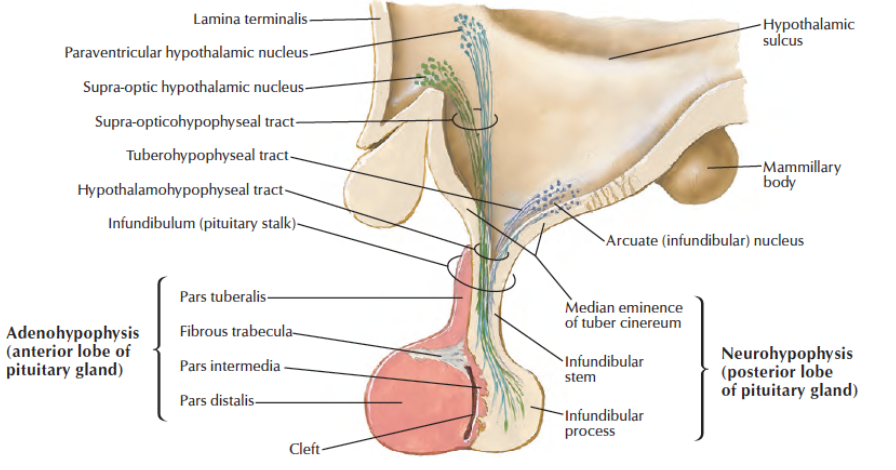
Neurohypophysis
1. Posterior lobe or neural lobe, pars posterior:
It is smaller than the anterior lobe and lies in the posterior concavity of the larger anterior lobe.
2. lnfundibular stem, which contains the neural connections of the posterior lobe with the hypothalamus.
3. Median eminence of the tubercinerium which is continuous with the infundibular stem

Arterial Supply
The hypophysis cerebri is supplied by the following branches of the internal carotid artery.
1. One superior hypophyseal artery on each side
2. One inferior hypophyseal artery on each side.
Each superior hypophyseal artery supplies:
a. Ventral part of the hypothalamus.
b. Upper part of the infundibulum.
c. Lower part of the infundibulum through a separate long descending branch, called the trabecular artery.
Each inferior hypophyseal artery divides into medial and lateral branches which join one another to form an arterial ring around the posterior lobe.
Branches from this ring supply the posterior lobe and also anastomose with branches from the superior hypophyseal artery.
The anterior lobe or pars distalis is supplied exclusively by portal vessels arising from capillary tufts formed by the superior hypophyseal arteries.
The long portal vessels drain the median eminence and the upper infundibulum, and the short portal vessels drain the lower infundibulum.
The portal vessels are of great functional importance because they carry the hormone releasing factors from the hypothalamus to the anterior lobe where they control the secretory cycles of different glandular cells.

Venous Drainage
Short veins emerge on the surface of the gland and drain into neighbouring dural venous sinuses.
The hormones pass out of the gland through the venous blood, and are carried to their target cells.
Hormones
Anterior lobe
Chromophilic cells 50%.
1. Acidophils/alpha-cells; about 43%
a. Somatotrophs: Secrete growth hormone (STH,GH).
b. Mammotrophs (prolactin cells): Secrete lactogenic hormone.
c. Corticotrophs: Secrete ACTH.
2. Basophils/beta-cells, about 7% of cells
a. Thyrotrophs: Secrete TSH.
b. Gonadotrophs: Secrete FSH.
c. Luteotrophs: Secrete LH or ICSH.
Chromophobic cells 50% represent the non-secretory phase of the other cell types, or their precursors.
Intermediate Lobe
It is made up of numerous basophil cells, and chromophobe cells surrounding masses of colloid material.
It secretes the melanocyte stimulating hormone (MSH).
Posterior Lobe
It is composed of:
1. A large number of nonmyelinated fibres hypothalamo- hypophyseal tract.
2. Modified neurological cells, called pituicytes. They have many dendrites which terminate on or near the
sinusoids.
Hypothalamo-hypophyseal portal system
The hypothalamo-hypophyseal tract begins in the preoptic and paraventricular nuclei of the hypothalamus.
Its short fibres terminate in relation to capillary tufts of portal vessels, providing the possibility for a neural control of the secretory activity of the anterior lobe.
The long fibres of the neurosecretory tract pass to the posterior lobe and terminate near vascular sinusoids.
The hormones related to the posterior lobe are:
a. Vasopressin (ADH) which acts on kidney tubules.
b. Oxytocin which promotes contraction of the uterine and mammary smooth muscle.
These hormones are actually secreted by the hypothalamus, from where these are transported through the
hypothalamo-hypophyseal tract to the posterior lobe of the gland.

Clinical Anatomy
Pituitary tumours give rise to two main categories of symptoms:
A. General symptoms due to pressure over surrounding structures:
a. The sella turcica is enlarged in size.
b. Pressure over the central part of optic chiasma causes bitemporal hemianopia
c. Pressure over the hypothalamus may cause one of the hypothalamic syndromes like obesity of Frolich's syndrome in cases with Rathke's pouch tumours.
d. A large tumour may press upon the third ventricle, causing a rise in intracranial pressure.
B. Specific symptoms depending on the cell type of the tumour.
a. Acidophil or eosinophil adenoma causes acromegaly in adults and gigantism in younger patients.
b. Basophil adenoma causes Cushing's syndrome.
c. Chromophobe adenoma causes effects of hypopituitarism.
d. Posterior lobe damage causes diabetes insipidus, although the lesion in these cases usually lies in the hypothalamus.
Watch Lectures on YouTube:
No comments:
Post a Comment