Internal ear or labyrinth lies in the petrous part of the temporal bone.
It consist of bony labyrinth within which there is a membranous labyrinth.
The membranous labyrinth is filled with a fluid called endolymph.
Membranous labyrinth is separated from the bony labyrinth by another fluid called perilymph
Bony Labyrinth
Consists of three parts.
- Cochlea anteriorly.
- Vestibule in the middle.
- Semicircular canals posteriorly
Cochlea
Bony cochlea resembles the shell of a common snail.
It forms the anterior part of labyrinth.
It has a conical central axis known as the modiolus around which the cochlear canal makes two and half turns.
The modiolus is directed forwards and laterally.
Its apex points towards the anterosuperior part of the medial wall of the middle ear.
Base is towards the fundus of the internal acoustic meatus
A spiral ridge of bone - spiral lamina, projects from the modiolus and
partially divides the cochlear canal into
- Scala vestibuli above and
- Scala tympani below
These relationships apply to the lowest part or basal turn of cochlea.
The division between the two passages is completed by the basilar membrane.
The scala vestibuli communicates with scala tympani at the apex if the cochlea by a small opening called the Helicotrema.
Vestibule:
- This is central part of the bony labyrinth.
- It lies medial to the middle ear cavity.
- Its lateral wall opens into the middle ear at the fenestra vestibuli, which is closed by the footplate of the stapes.
- Three semicircular canals opens its posterior wall.
- The medial wall is related to the internal acoustic meatus and presents
Spherical recess in front &
Elliptical recess behind.
- The two recess are separated by a vestibular crest which splits inferiorly to enclose the cochlear recess
Just below the elliptical recess, there is the opening of a diverticulum, the aqueduct of the vestibule which opens at a narrow fissure on the posterior aspect of the petrous temporal bone, posterolateral to the internal acoustic meatus.
It is plugged in life by ductus endolymphaticus and a vein; no perilymph escapes through it
Semicircular Canals
There are three bony semicircular canals
1. Anterior or superior
2. Posterior
3 Lateral.
Each canal has two ends.
They lie posterosuperior to the vestibule
They are set at right angles to each other.
Each canal is 2/3rd of a circle.
One end is dilated at one end to form ampulla
Three canals open into the vestibule by five openings
Anterior or Superior Semicircular canals
It lie in a vertical plane at right angles to the long axis of the petrous temporal bone.
It is convex upwards.
Its position is indicated by the arcuate eminence seen on the anterior surface of the petrous temporal bone.
Its ampulla is situated anterolaterally.
Its posterior end unites with the upper end of the posterior canal to form crus commune, which opens into the medial wall of the vestibule.
Posterior Semicircular canal:
It lies in a vertical plane parallel to the long axis of petrous temporal bone.
It is convex backwards.
Its ampulla lies at its lower end.
The upper end joins the anterior canal to form crus commune
Lateral Semicircular Canal:
It lies in the horizontal plane.
Its convexity directed posterolaterally.
Its ampulla lies anteriorly, close to the ampulla of the anterior canal.
- The lateral semicircular canals of two sides lie in the same plane.
- The anterior canal of one side lie in the plane of the posterior canal of the other side
Membranous Labyrinth:
It is a complicated, but continuous closed cavity filled with Endolymph.
The epithelium of membranous labyrinth is specialised to form receptors for sound - The organ of Corti
For static balance - the maculae.
For kinetic balance - the cristae
The membranous labyrinth consists of three main parts:
1. Spiral duct of cochlea or organ of Corti, anteriorly.
2. The utricle and saccule - the organs of Static balance within the vestibule.
3. The semicircular ducts with Cristae - the organ of kinetic balance, posteriorly
Duct of the cochlea or the Scala media:
Spiral duct occupies the middle part of the cochlear canal between the Scala vestibuli and scala tympani.
It is triangular in cross-section.
The floor is formed by the basilar membrane.
The roof is formed by vestibular or Reissner’s membrane
The outer wall by the bony wall of the cochlea.
The Basilar membrane supports the spiral organ of Corti, which is the end organ for hearing.
It comprises rods of Corti and hair cells
Hair is embedded in a gelatinous membrane called the membrana tectoria.
The organ of Corti is innervated by peripheral processes of the bipolar cells located in the spiral ganglion.
This ganglion is located in the spiral canal present within the modiolus at the base of the spiral lamina.
The Central processes form the cochlear nerve
Posteriorly the duct of cochlea is connected to the saccule by a narrow ductus reunions.
The sound waves reaching the endolymph through the vestibular membrane
Make appropriate parts of the basilar membrane vibrate.
So that different parts of organ of Corti are stimulated by different frequencies of sound.
The loudness of the sound depends on the amplitude of vibration
Saccule:
It lies in the anteroinferior part of the vestibule.
It is connected to the basal turn of cochlear duct by ductus reunions
Utricle:
It is larger than saccule.
It lies in the posterosuperior part of the vestibule.
It receives the ends of three semicircular ducts through five openings.
The duct of saccule unites with the duct of the utricle to form ductus endolymphaticus.
The ductus endolymphaticus ends in a dilatation, the saccus endolymphaticus.
The ductus and saccus occupy the aqueduct of the vestibule
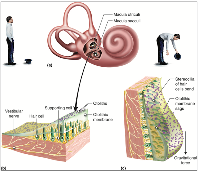
The medial walls of the saccule and utricle are thickened to form a macula in each chamber.
The maculae are end organs that give information about the position of the head.
They are static balance receptors.
They are supplied by peripheral processes of neutrons in the vestibular ganglion.
Saccule get stimulated by vertical linear motions like going in lift.
Utricle gets stimulates by horizontal linear motions like going in car.
Semicircular ducts:
They lie within the corresponding bony canals.
Each duct has an ampulla corresponding to that of bony canal.
It each ampulla, there is an end organ called ampullary crest or crista or cupola.
Cristae respond to pressure changes in the endolymph caused by movements of the head.
Blood Supply of Labyrinth:
- The arterial supply is mainly by labyrinthine branch of basilar artery, which accompanies the vestibulocochlear nerve.
- Partly by the stylomastoid branch of posterior auricular artery
- Labyrinthine veins drains into the superior petrosal sinus or transverse sinus.
- Other inconstant veins emerge at different points and open separately into superior and inferior petrosal sinuses and the internal jugular vein.
Development:
1. External auditory meatus - Dorsal part of 1st ectodermal cleft.
2. Auricle - tubercle appearing on the 1st and 2nd branchial arches around the opening of external auditory meatus
3. Middle ear cavity and auditory tube - tubotympanic recess.
4. Ossicles
A. Malleus and incus - from 1st arch cartilage
B. Stapes - from 2nd arch cartilage.
5. Muscles.
a. Tensor tympani - from 1st pharyngeal arch mesoderm
b. Stapedius - from 2nd pharyngeal arch mesoderm
6. Membranous labyrinth from ectodermal vesicle on each side of hindbrain vesicle.
Organ of Corti - ectodermal
Clinical Anatomy:
Endolymph is produced by striae vascularis. This process requires melanocytes. The disorders of melanocytes, i.e. albinism, are associated with deafness.
Acoustic neuroma is a tumour of Schwann cells of 8th nerve. If neuroma extends into internal auditory meatus, the 7th nerve will get compressed. There will be both 7th and 8th nerve paralysis.
Watch lectures on YouTube:
Internal Ear - The Bony Labyrinth | Parts | Features | Scala vestibuli | Scala Tympani | Perilymph


No comments:
Post a Comment