Monday, June 29, 2020

Maxillary Artery

This is the larger terminal branch of the external carotid artery,
given off behind the neck of the mandible

It has a wide territory of distribution, and supplies

  • 1. The external and middle ears, and the auditory tube
  • 2. The dura mater
  • 3. The upper and lower jaws and teeth
  • 4. The muscles of the temporal and infratemporal regions
  • 5. The nose and paranasal air sinuses
  • 6. The palate
  • 7. The root of the pharynx.

COURSE AND RELATIONS

For descriptive purposes, the maxillary artery is divided into three parts.

  • I. The first (mandibular) part runs horizontally forwards,first between the neck of the mandible and the sphenomandibular ligament, below the auriculotemporal nerve, and then along the lower border of the lateral pterygoid.
  • 2. The second (pterygoid) part runs upwards and forwards superficial to the lower head of the lateral pterygoid.

  • 3. The third (pterygopalatine) part passes between the two heads of the lateral pterygoid and through the pterygomaxillary fissure, to enter the pterygopalatine fossa. 

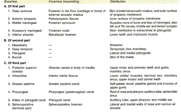
BRANCHES OF FIRST PART OF THE MAXILLARY ARTERY

  • 1. deep auricular artery
  • 2. anterior tympanic branch
  • 3. middle meningeal artery
  • 4. accessory meningeal artery
  •  5. inferior alveolar artery 

BRANCHES OF SECOND PART OF THE MAXILLARY ARTERY

mainly muscular

  • 1. masseteric,
  • 2. deep temporal branches - Anterior & Posterior
  • 3. to the pterygoid muscles 
  • 4. buccal branch 

BRANCHES OT THIRD PART OF THE MAXILLARY ARTERY

  • 1. posterior superior alveolar artery 
  • 2. infraorbital artery
  • 3. greater palatine artery
  • 4. pharyngeal branch
  • 5. artery of the pterygoid canal 
  • 6. sphenopalatine artery 


Watch Lectures on YouTube:


Sunday, June 28, 2020

Muscles of Mastication

Masseter

Quadrilateral muscle

covers the lateral surface of ramus of mandible

Origin

1. Superficial Layer: anterior 2/3 of lower border of zygomatic arch

2. Deep layer: deep surface of zygomatic arch

3. Middle Layer: posterior 1/3 of lower border of zygomatic arch

Insertion

Superficial layer into lower part of lateral surface of ramus of mandible

Deep Layer into rest of the ramus Middle layer into central part of ramus

Nerve supply: Masseteric Nerve, branch of anterior division of mandibular Nerve

Action:

  • Elevation of mandible
  • Superficial fibres cause protrusion

Temporalis

  • fan shaped muscle
  • fills the temporal fossa

Origin:

  • temporal fossa including zygomatic bone
  • temporal fascia

Insertion:

  • Margins & deep surface of coronoid process of mandible
  • anterior border of ramus of mandible

Nerve Supply: Two deep temporal nerve from anterior division of mandibular nerve

Action:

  • elevate the mandible
  • helps in side to side grinding movements 
  • retracts the protruded mandible 

Lateral Pterygoid

Origin:
Upper head
- from infra temporal surface and crest of greater wing of sphenoid

Lower head - from lateral surface of lateral pterygoid plate

Insertion:

  • pterygoid fovea on the anterior surface of neck of the mandible
  • anterior margin of the articular disc & capsule of temporomandibular joint

Nerve supply: branch from anterior division of mandibular nerve

Action:

  • depression of mandible
  • lateral & medial pterygoids protrudes the mandible
  • right lateral & medial pterygoids turn the chin to left side, as a part of grinding movement 

RELATIONS OF LATERAL PTERYGOID

The lateral pterygoid may be regarded as the key muscle of this region because its relations provide a fair idea about the layout of structures in the infratemporal fossa.
The relations are as follows:
Superficial
1. Masseter
2. Ramus of the mandible
3. Tendon of the temporalis
4. The maxillary artery


Deep
1. Mandibular nerve
2. Middle meningeal artery.
3. Sphenomandibular ligament
4. Deep head of the medial pterygoid. 


Structures Emerging at the Upper Border

1. Deep temporal nerves
2. Masseteric nerve.

Structures Emerging at the lower Border
1. Lingual nerve
2. Inferior alveolar nerve
3. The middle meningeal artery passes upwards deep to it

Structures Passing through the Gap Between the Two Heads
1. The maxillary artery enters the gap
2. The buccal branch of the mandibular nerve comes out through the gap
The pterygoid plexus of veins surrounds the lateral pterygoid. 

Medial Pterygoid Origin

Superficial head: from tuberosity of maxilla & adjoining bone

Deep head: from medial surface of lateral pterygoid plate & adjoining process of palatine bone

Insertion: roughened area on the medial surface of the angle & adjoining ramus of mandible

Nerve Supply: Nerve to medial pterygoid, a branch from the main trunk of mandibular nerve

Action:

  • elevation of mandible protrusion of mandible
  • right medial & right lateral pterygoid moves the chin to left side 

RELATIONS OF MEDIAL PTERYGOID

The superficial and deep heads of medial pterygoid enclose the lower head of lateral pterygoid muscle
Superficial Relations

The upper part of the muscle is separated from the lateral pterygoid muscle by:

  • 1. The lateral pterygoid plate
  • 2. The lingual nerve
  • 3. The inferior alveolar nerve.

Lower down the muscle is separated from the ramus of the mandible by

  • 1. the lingual and inferior alveolar nerves,
  • 2. the maxillary artery, and
  • 3. the sphenomandibular ligament.
Deep Relations
The relations are:
  • 1. Tensor veli palatini
  • 2. Superior constrictor of pharynx
  • 3. Styloglossus
  • 4. Stylopharyngeus attached to the styloid process 

Watch lectures on YouTube:


Friday, June 26, 2020

Temporal & Infratemporal Fossa

Temporal Fossa

lies on the side of the skull,

BOUNDARIES

  • Anterior: Zygomatic and frontal bones. 
  • Posterior: Inferior temporal line and supramastoid crest.
  • Superior: Superior temporal line 
  • lnferior : Zygomatic arch.
  • Floor: Parts of frontal, parietal, temporal and greater wing of sphenoid.

  • Temporalis muscle is attached to the floor and inferior temporal line.

CONTENTS

1. Temporalis muscle.
2. Middle temporal artery (branch of superficial temporal artery
3. Zygomaticotemporal nerve and artery.
4. Deep temporal nerves for supplying temporalis muscle.
5. Deep temporal artery, branch of maxillary artery.


Infratemporal Fossa

It is an irregular space below zygomatic arch.

BOUNDARIES

  • Anterior: Posterior surface of body of maxilla. 
  • Roof: Infratemporal surface of greater wing of sphenoid.
  • Medial: Lateral pterygoid plate and pyramidal process of palatine bone.
  • Lateral: Ramus of mandible

Contents
1. Lateral pterygoid muscle.
2. Medial pterygoid muscle
3. Mandibular nerve with its branches.
4. Maxillary nerve with posterior superior alveolar nerve.

5. Chorda tympani, branch of VII nerve.
6. 1st and 2nd parts of maxillary artery with their branches.
7. Posterior superior alveolar artery, branch of 3rd part of maxillary artery.
8. Accompanying veins 


Watch Lectures on YouTube:


Parotid Gland

  • The parotid is the largest of the salivary glands.
  • It weighs about 15 g.
  • It is situated below the external acoustic meatus, between the ramus of the mandible and the sternocleidomastoid. 
  • The gland overlaps these structures.
  • Anteriorly, the gland also overlaps the masseter muscle.
  • A part of this forward extension is often detached, and is known as the accessory parotid, and it lies between the zygomatic arch and the parotid duct.

Capsule of Parotid Gland

  • The investing layer of the deep cervical fascia forms a capsule for the gland.

  • The fascia splits (between the angle of the mandible and the mastoid process) to enclose the gland.
  • The superficial lamina, thick and adherent to the gland, is attached above to the zygomatic arch.

  • The deep lamina is thin and is attached to the styloid process, the angle and posterior border of the ramus of the mandible and the tympanic plate.
  • A portion of the deep lamina, extending between the styloid process and the mandible, is thickened to form the stylomandibular ligament which separates the parotid gland from the submandibular salivary gland.
  • The ligament is pierced by the external carotid artery

Clinical Anatomy

  • Parotid swellings are very painful due to the unyielding nature of the parotid fascia.
  • Mumps is an infectious disease of the salivary glands (usually the parotid) caused by a specific virus,
  • Viral parotitis or mumps characteristically does not suppurate. Its complications are orchitis and pancreatitis 

External Features

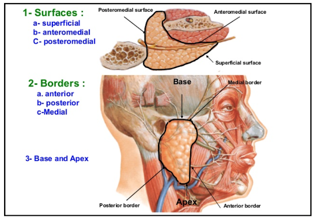
The gland resembles a three sided pyramid. The apex of the pyramid is directed downwards The gland has four surfaces:

  • a. Superior (base of the pyramid)
  • b. Superficial
  • c. Anteromedial
  • d. Posteromedial.
The surfaces are separated by three borders

  • a. Anterior
  • b. Posterior
  • c. Medial/pharyngeal 

Relations

  • The apex overlaps the posterior belly of the digastric and the adjoining part of the carotid triangle.

  • The cervical branch of the facial nerve and the two divisions of the retromandibular vein emerge near the apex. 

Surfaces

The superior surface or base forms the upper end of the gland which is small and concave.
It is related to:

  • a. The cartilaginous part of the external acoustic meatus.
  • b. The posterior surface of the temporomandibular joint.
  • c. The superficial temporal vessels. 
  • d. The auriculotemporal nerve 

The superficial surface is the largest of the four surfaces.
It is covered with:

  • a. Skin
  • b. Superficial fascia containing the anterior branches of the great auricular nerve, the preauricular or superficial parotid lymph nodes and the posterior fibres of the platysma and risorius.
  • c. The parotid fascia which is thick and adherent to the gland.
  • d. A few deep parotid lymph nodes embedded in the gland 

The anteromedial surface is grooved by the posterior border of the ramus of the mandible. It is related to:

  • a. The masseter
  • b. The lateral surface of the temporomandibular joint.
  • c. The posterior border of the ramus of the mandible
  • d. The medial pterygoid
  • e. The emerging branches of the facial nerve. 

The posteromedial surface is moulded to the mastoid and the styloid processes and the structures attached to them.

Thus it is related to:

  • a. The mastoid process, with the sternocleidomastoid and the posterior belly of the digastric.
  • b. The styloid process, with structures attached to it.
  • c. The external carotid artery enters the gland through this surface and the internal carotid artery lies deep to the styloid process 

Borders

  • The anterior border separates the superficial surface from the anteromedial surface.

  • It extends from the anterior part of the superior surface to the apex.

The following structures emerge at this border: 

  • a. The parotid duct.
  • b. Most of the terminal branches of the facial nerve.
  • c. The transverse facial vessels. In addition, the accessory parotid gland lies on the parotid duct close to this border.

The posterior border separates the superficial surface from the posteromedial surface. 

  • It overlaps the sternocleidomastoid.

The medial edge or pharyngeal border separates the anteromedial surface from the posteromedial surface.

  • It is related to the lateral wall of the pharynx 

Structures within the parotid gland

From medial to the lateral side, these are as follows. 

Arteries

  • The external carotid artery enters the gland through its posteromedial surface.
  • The maxillary artery leaves the gland through its anteromedial surface.
  • The superficial temporal artery gives transverse facial artery and emerges at the anterior part of the superior surface. 

Veins: 

  • The retromandibular vein is formed within the gland by the union of the superficial temporal and maxillary veins. 
  • In the lower part of the gland, the vein divides into anterior and posterior divisions which emerge close to the apex (lower pole) of the gland. 

Nerve: 

  • facial nerve exits from cranial cavity through stylomastoid foramen and enters the gland through the upper part of its posteromedial surface, and divides into its terminal branches within the gland.
  • The branches leave the gland through its anteromedial surface, and appear on the surface at the anterior border. 
  • Facial nerve lies in relation to isthmus of the gland which separates large superficial part from small deep part of the gland.

Facial nerve divides into two branches

  • a. Temporofacial: Divides into temporal and zygomatic branches.
  • b. Cervicofacial: Divides into buccal, marginal mandibular and cervical branches.
  • The various branches (5-6) of facial nerve radiate like a goose-foot from the curved anterior border of the parotid gland to supply the respective muscles of facial expression. This pattern of branching is called "pes anserinus"
Lymph Nodes: Parotid lymph nodes. 

Patey's faciovenous plane

The gland is composed of a large superficial and a small deep part the two being connected by an 'isthmus' around which facial nerve divides 

Accessory processes of parotid gland

  • Facial process - along parotid duct 
  • Pterygoid process - between mandibular ramus and medial pterygoid.
  • Glenoid process - between external acoustic meatus and temporomandibular joint 
  • Poststyloid process 

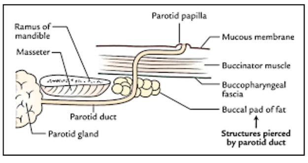
Parotid Duct / Stenson’s Duct

  • It is thick walled and is about 5 cm long.
  • It emerges from the middle of the anterior border of the gland
  • It runs forwards and slightly downwards on the masseter. Here its relations are:
Superiorly
1. Accessory parotid gland.
2. The transverse facial vessels.
3. Upper buccal branch of the facial nerve.
Inferiorly
The lower buccal branch of the facial nerve.

At the anterior border of the masseter, the parotid duct turns medially and pierces:
a. The buccal pad of fat.
b. The buccopharyngeal fascia.
c. The buccinator (obliquely). 

Because of the oblique course of the duct through the buccinator, inflation of the duct is prevented during blowing.
The duct runs forwards for a short distance between the buccinator and the oral mucosa. 

Finally, the duct turns medially and opens into the vestibule of the mouth (gingivobuccal vestibule) opposite the crown of the upper second molar tooth 

Clinical Anatomy

  • Parotid abscess may be caused by spread of infection from the opening of parotid duct in the mouth cavity.
  • Parotidectomy is the removal of the parotid gland.

  • After this operation, at times, there may be regeneration of the secretomotor fibres in the auriculotemporal nerve which join the great auricular nerve. 
  • This causes stimulation of the sweat glands and hyperaemia in the area of its distribution, thus producing redness and sweating in the area of skin supplied by the nerve. 
  • This clinical entity is called Frey Syndrome.
  • Whenever, such a person chews there is increased sweating in the region supplied by auriculotemporal nerve.
  • It is also called 'auriculotemporal syndrome'

BIood Supply

  • The parotid gland is supplied by the external carotid artery and its branches that arise within the gland. 
  • The veins drain into the external jugular vein and internal jugular vein. 

Nerve Supply

  • 1. Parasympathetic nerves are secretomotor They reach the gland through the auriculotemporal nerve.
  • 2 Sympathetic nerves are vasomotor, and are derived from the plexus around the middle meningeal artery.
  • 3 Sensory nerves to the gland come from the auriculotemporal nerve, but the parotid fascia is innervated by the sensory fibres of the great auricular nerve (C2, C3) 


Lymphatic Drainage

Lymph drains first to the parotid nodes and from there to the upper deep cervical nodes.

DEVELOPMENT

The parotid gland is ectodermal in origin.
It develops from the buccal epithelium just lateral to the angle of mouth. The outgrowth branches repeatedly to form the duct system and acini.
The
mesoderm forms the intervening connective tissue septa. 

Clinical Anatomy

  • A parotid abscess is best drained by horizontal incision known as Hilton's method below the angle of mandible
  • During surgical removal of the parotid gland or parotidectomy the facial nerve is preserved by removing the gland in two parts, superficial and deep separately, 
  • The plane of cleavage is defined by tracing the nerve from behind forwards.
  • Mixed parotid tumour is a slow growing lobulated painless tumour without any involvement of the facial nerve.
  • Malignant change of such a tumour is indicated by pain, rapid growth, fixity with hardness, involvement of the facial nerve, and enlargement of cervical lymph nodes. 
  • The parotid calculi may get formed within the parotid gland or in its Stenson's duct. 
  • These can be located by injecting a radiopaque dye through its opening in the vestibule of the mouth. The procedure is called 'Sialogram'
  • The duct can be examined by a spatula or bidigital examination. 


Watch Lectures on YouTube:


Thursday, June 25, 2020

Infrahyoid Muscles / Ribbon Muscles

The infrahyoid muscles are: 

  • a. Sternohyoid
  • b. Sternothyroid
  • c. Thyrohyoid
  • d. Omohyoid.

These ribbon muscles have the following general features.

  • a. They are arranged in two layers, superficial (sternohyoid and omohyoid) and deep (sternothyroid and thyrohyoid).
  • b. All of them are supplied by the ventral rami of first, second and third cervical spinal nerves.
  • c. Because of their attachment to the hyoid bone and to the thyroid cartilage, they move these structures. 
  • d. Sternohyoid, superior belly of omohyoid, sternothyroid lie superficial to the lateral or superficial convex surface of the thyroid gland.
  • e. The anterior surface of isthmus of thyroid gland is covered by right and left sternothyroid and sternohyoid muscles.

Watch lectures on YouTube:


Tuesday, June 23, 2020

Ansa Cervicalis

  • This is a thin nerve loop that lies embedded in the anterior wall of the carotid sheath over the lower part of the laryrrx.
  • It supplies the infrahyoid muscles
Formation
It is formed by a superior and an inferior root.
  • The superior root is the continuation of the descending branch of the hypoglossal nerve.
    Its fibres are derived from the first cervical nerve. This root descends over the internal carotid artery and the common carotid artery.


  • The inferior root or descending cervical nerve is derived from second and third cervical spinal nerves.
    As this root descends, it winds round the internal jugular vein, and then continues anteroinferiorly to join the superior root in front of the common carotid artery

Distribution

  • Superior root: To the superior belly of the omohyoid.
  • Ansa cervicalis: To the sternohyoid, the sternothyroid and the inferior belly of the omohyoid.

  • Note that the thyrohyoid and geniohyoid are supplied by separate branches from the first cervical nerve through the hypoglossal nerve 

Watch the lectures on YouTube:


Monday, June 22, 2020

Arteries of Carotid Triangle

Common Carotid Artery

  • The right common carotid artery is a branch of the brachiocephalic artery.
    It begins in the neck behind the right sternoclavicular joint.
  • It begins in the neck behind the right sternoclavicular joint.
  • The left common carotid artery is branch of the arch of the aorta.
    It begins in the thorax in front of the trachea opposite a point a little to the left of the centre of the manubrium.
    It ascends to the back of left sternoclavicular joint and enters the neck.


  • In the neck, both arteries have a similar course. Each artery runs upwards within the carotid sheath, under cover of the anterior border of the sternocleidomastoid.
    It lies in front of the lower four cervical transverse processes.
    At the level of the upper border of the thyroid cartilage, the artery ends by dividing into the external and internal carotid arteries
  • It lies in front of the lower four cervical transverse processes.
  • At the level of the upper border of the thyroid cartilage, the artery ends by dividing into the external and internal carotid arteries

Carotid Sinus

  • The termination of the common carotid artery, or the beginning of the internal carotid artery shows a slight dilatation, known as the carotid sinus.
  • In this region, the tunica media is thin,
  • but the adventitia is relatively thick and receives a rich innervation from the glossopharyngeal and sympathetic nerves.
  • The carotid sinus acts as a baroreceptor of pressure receptor and regulates blood pressure 


Carotid Body

  • Carotid body is a small, oval reddish brown structure
  • situated behind the bifurcation of the common carotid artery.
  • It receives a rich nerve supply mainly from the glossopharyngeal nerve, but also from the vagus and sympathetic nerves.
  • It acts as a chemoreceptor and responds to changes in the oxygen, carbon dioxide and pH content of the blood.
  • Other allied chemoreceptors are found near the arch of the aorta, the ductus arteriosus, and the right subclavian artery. These are supplied by the vagus nerve. 

Clinical Anatomy

  • The carotid sinus is richly supplied by nerves.
  • In some persons, the sinus may be hypersensitive. In such persons, sudden rotation of the head may cause slowing of heart. This condition is called as "carotid sinus syndrome".
    The supraventricular tachycardia may be controlled by carotid sinus massage/ due to inhibitory effects of vagus nerve on the heart.
    The necktie should not be tied tightly, as it may compress both the internal carotid arteries, supplying the brain. 
  • The supraventricular tachycardia may be controlled by carotid sinus massage/ due to inhibitory effects of vagus nerve on the heart.
  • The necktie should not be tied tightly, as it may compress both the internal carotid arteries, supplying the brain. 

External Carotid Artery

Course & Relations

  • The external carotid artery begins in the carotid triangle at the level of the upper border of the thyroid cartilage opposite the disc between the third and fourth cervical vertebrae.
  • It runs upwards and slightly backwards and laterally, and terminates behind the neck of the mandible by dividing into the maxillary and superficial temporal arteries.
    The external carotid artery has a slightly curved course, so that it is anteromedial to the internal carotid artery in its lower part, and anterolateral to the internal carotid artery in its upper part.
  • ln the carotid triangle, the external carotid artery is comparatively superficial, and lies under cover of the anterior border of the sternocleidomastoid.
    The artery is crossed superficially by the cervical branch of the facial nerve, the hypoglossal nerve, and the facial, lingual and superior thyroid veins. 
  • The artery is crossed superficially by the cervical branch of the facial nerve, the hypoglossal nerve, and the facial, lingual and superior thyroid veins. 

Deep to the artery, there are:
a. The wall of the pharynx.
b. The superior laryngeal nerve which divides into the external and internal laryngeal nerves.
c. The ascending pharyngeal artery


Above the carotid triangle, the external carotid artery lies deep in the substance of the parotid gland. Within the gland, it is related superficially to the retromandibular vein and the facial nerve


Deep to the external carotid artery, there are:
a. The internal carotid artery.
b. Structures passing between the external and internal carotid arteries; these being styloglossus, stylopharyngeus, IX nerve, pharyngeal branch of X, and styloid process.
c. Two structures deep to the internal carotid artery, namely the superior laryngeal nerve and the superior cervical sympathetic ganglion 

Branches

The external carotid artery gives off eight branches which may be grouped as follows.
Anterior

1. Superior thyroid

2. Lingual
3. Facial
Posterior
1. Occipital
2. Posterior auricular. 

Medial

1. Ascending pharyngeal

Terminal

1. Maxillary
2. Superficial temporal 

Superior Thyroid Artery

  • arises from the external carotid artery just below the level of the greater cornua of the hyoid bone. runs downwards and forwards parallel and just superficial to the external laryngeal nerve.

  • passes deep to the three long infrahyoid muscles to

  • reach the upper pole of the lateral lobe of the thyroid gland.
  • relationship to the external laryngeal nerve, which supplies the cricothyroid muscle is important to the surgeon during thyroid surgery.
  • The artery and nerve are close to each other higher up, but diverge slightly near the gland.
  • To avoid injury to the nerve, the superior thyroid artery is ligated as near the gland as possible
  • Apart from its terminal branches to the thyroid gland, it gives one important branch, the superior laryngeal artery which pierces the thyrohyoid membrane in company with the internal laryngeal nerve
  • The superior thyroid artery also gives a sternocleidomastoid branch to that muscle and a cricothyroid branch that anastomoses with the artery of the opposite side in front of the cricovocal membrane. 

Lingual Artery

  • arises from the external carotid artery opposite the tip of the greater cornua of the hyoid bone.

  • It is tortuous in its course.

  • Its course is divided into three parts by the hyoglossusmuscle.

  • The first part lies in the carotid triangle.

  • It forms a characteristic upward loop which is crossed by the hypoglossal nerve.

  • The lingual loop permits free movements of the hyoid bone.

  • The second part lies deep to the hyoglossus along the upper border of hyoid bone.

  • It is superficial to the middle constrictor of the pharynx.

  • The third part is called the arteria profunda linguae, or the deep lingual artery.

  • It runs upwards along the anterior border of the hyoglossus, and then hoizontally forwards on the undersurface of the tongue as the fourth part.

  • In its vertical course, it lies between the genioglossus medially and the inferior longitudinal muscle of the tongue laterally.

  • The horizontal part of the artery is accompanied by the lingual nerve.

  • During surgical removal of the tongue, the first part of the artery is ligated before it gives any branch to the tongue or to the tonsil. 

Facial Artery:

  • arises from the external carotid just above the tip of the greater cornua of the hyoid bone.
  • It runs upwards first in the neck as cervical part and then on the face as facial part. The course of the artery in both places is tortuous.
    The tortuosity in the neck allows free movements of the pharynx during deglutition. On the face, it allows free movements of the mandible, the lips and the cheek during mastication and during various facial expressions.
    The artery escapes traction and pressure during these movements.

  • The tortuosity in the neck allows free movements of the pharynx during deglutition. On the face, it allows free movements of the mandible, the lips and the cheek during mastication and during various facial expressions.
  • The artery escapes traction and pressure during these movements.
  • The cervical part of the facial artery runs upwards on the superior constrictor of pharynx deep to the posterior belly of the digastric, with the stylohyoid and to the ramus of the mandible.
    It grooves the posterior border of the submandibular salivary gland.
    Next the artery makes an S-bend (two loops) first winding down over the submandibular gland, and then up over the base of the mandible

  • The facial part of the facial artery enters the face at anteroinferior angle of masseter muscle, runs upwards close to angle of mouth, side of nose till medial angle of eye. 

The cervical part of the facial artery gives off 

1. ascending palatine,
2. tonsillar,
3. submental, and 

4. glandular branches for the submandibular salivary gland and lymph nodes.

  • The ascending palatine artery arises near the origin of the facial artery. It passes upwards between the styloglossus and the stylopharyngeus, crosses over the upper border of the superior constrictor and supplies the tonsil and the root of the tongue.
  • The submental branch is a large artery which accompanies the mylohyoid nerve, and supplies the submental triangle and the sublingual salivary gland 

The facial part

  • It enters the face by winding around the base of the mandible, and by piercing the deep cervical fascia, at the anteroinferior angle of the masseter muscle.
  • It can be palpated here and is called ‘anaesthetist's artery'.
  • First it runs upwards and forwards to a point 1.25 cm lateral to the angle of the mouth.
  • Then it ascends by the side of the nose up to the medial angle of the eye, where it terminates by supplying the lacrimal sac; and by anastomosing with the dorsal nasal branch of the ophthalmic artery.
  • The facial artery is very tortuous.
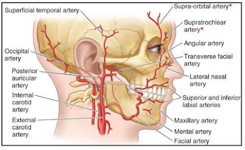
  • The tortuosity of the artery prevents its walls from being unduly stretched during movements of the mandible, the lips and the cheeks.
  • It lies between the superficial and deep muscles of the face.

Branches

  • The anterior branches on the face are large and named.
    They are:
1. lnferior labial, to the lower lip.
2.
Superior labial, to the upper lip and the anteroinferior part of the nasal septum.
3.
Lateral nasal, to the ala and dorsum of the nose.
  • The posterior branches are small and unnamed.
Anastomoses
1. The large anterior branches anastomose with similar branches of the opposite side and with the mental artery. In the lips, anastomoses are large, so that cut arteries spurt from both ends
2. Small posterior branches anastomose with the transverse facial and infraorbital arteries.
3. At the medial angle of the eye, terminal branches of the facial artery anastomose with branches of the ophthalmic artery. This is, therefore, a site for anastomoses between the branches of the external and internal carotid arteries 

Occipital Artery

  • The occipital artery arises from the posterior aspect of the external carotid artery, opposite the origin of the facial artery
  • It is crossed at its origin by the hypoglossal nerve.
  • In the carotid triangle, the artery gives two sternocleidomastoid branches.
  • The upper branch accompanies the accessorynerve, and the lower branch arises near the origin of the occipital artery 

Posterior Auricular Artery

  • The posterior auricular artery arises from the posterior aspect of the external carotid just above the posterior belly of the digastric.
  • It runs upwards and backwards deep to the parotid gland, but superficial to the styloid process.
  • It crosses the base of the mastoid process, and ascends behind the auricle.
  • It supplies the back of the auricle, the skin over the mastoid process, and over the back of the scalp.
  • It is cut in incisions for mastoid operations.
  • Its stylomastoid branch enters the stylomastoid foramen, and supplies the middle ear, the mastoid antrum and air cells, the semicircular canals, and the facial nerve. 

Ascending Pharyngeal Artery

  • This is a small branch that arises from the medial side of the external carotid artery.
  • It arises very close to the lower end of external carotid artery.
  • It runs vertically upwards between the side wall of the pharynx, and the tonsil, medial wall of the middle ear and, the auditory tube.
  • It sends meningeal branches into the cranial cavity through the foramen lacerum, the jugular foramen and the hypoglossal canal 

Maxillary Artery

  • This is the larger terminal branch of the external carotid artery.
  • It begins behind the neck of the mandible under cover of the parotid gland.
  • It runs forwards deep to the neck of the mandible below the auriculotemporal nerve, and enters the infratemporal fossa 

Superficial Temporal Artery

1. It is the smaller terminal branch of the extemal carotid artery.
It begins, behind the neck of the mandible under cover of the parotid gland.

2. It runs vertically upwards, crossing the root of the zygoma or preauricular point, where its pulsations can be easily felt.
About 5 cm above the zygoma, it divides into anterior and posterior branches which supply the temple and scalp.

The anterior branch anastomoses with the supraorbital and supratrochlear branches of the ophthalmic artery.
3. In addition to the branches which supply the temple, the scalp, the parotid gland, the auricle and the facial muscles,

the superficial temporal artery gives off a transverse facial artery, and a middle temporal artery which runs on the temporal fossa deep to the temporalis muscle 


Watch lectures on YouTube:
Ascending Pharyngeal Artery | Branches | Distribution

Superior Thyroid Artery | Course | Branches | Distribution

Lingual Artery - Parts | Course | Relations | Branches

Facial Artery | Parts | Course | Relations | Branches & Distribution

Occipital artery | Course | Relations | Branches & Distribution

Posterior Auricular artery | Course | Relations | Branches & Distribution