Thursday, July 30, 2020

Prevertebral Muscles

Longus colli (cervicis)

Origin
a. The upper oblique part is from the anterior tubercles of the transverse processes of cervical vertebrae 3, 4, 5
b. Lower oblique part is from bodies of upper 2-3 thoracic vertebrae
c. Middle vertical part is from bodies of upper 3 thoracic and lower 3 cervical vertebrae

lnsertion 
a. Upper oblique part is into the anterior tubercle of the atlas
b. Lower oblique part is into the anterior tubercles of the transverse processes of 5th and 6th cervical vertebrae
c. Middle vertical part is into bodies of 1, 2,3,4 cervical vertebrae

Nerve supply
Ventral rami of nerves C3-C8

Actions
a. Flexes the neck
b. Oblique parts flex the neck laterally
c. Lower oblique part rotates the neck to the opposite side


Longus capitis
Origin
Anterior tubercles of transverse processes of cervical 3-6 vertebrae

Insertion
lnferior surface of basilar part of occipital bone

Nerve Supply
Ventral rami of nerves C1-C3

Action
Flexes the head


Rectus capitis anterior
Origin
Anterior surface of lateral mass of atlas in front of the occipital condyle
Insertion
Basilar part of the occipital bone
Nerve Supply
Ventral ramus of nerve C1
Action
Flexes the head


Rectus capitis lateralis
Origin
Upper surface of transverse process of atlas
Insertion
lnferior surface of jugular process of the occipital bone
Nerve Supply
Ventral rami of C2, C1 nerves ,
Action
Flexes the head laterally


SCALENOVERTEBRAL TRIANGLE
The triangle is present at the root of the neck.

Boundaries
Medial: Lower oblique part of longus colli
Lateral: Scalenus anterior
Apex: Transverse process of cervical C6 vertebra
Base: 1st part of subclavian artery
Posterior wall: Transverse process of C7, Ventral ramus of C8 nerve, neck of 1st rib and cupola of 
pleurae
Contents: 1st part of vertebral artery, cervical part
of sympathetic trunk


Watch the lectures on YouTube:


Wednesday, July 29, 2020

Styloid Apparatus

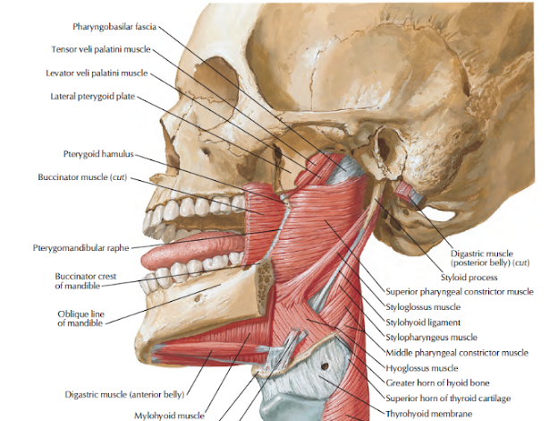
  • The styloid process with its attached structures is called the styloid apparatus. 
  • The structures attached to the process are three muscles and two ligaments. 
  • The muscles are the stylohyoid, styloglossus and stylopharyngeus
  • and ligaments are the stylohyoid and stylomandibular


  • The apparatus is of diverse origin. 
  • The styloid process/ the stylohyoid ligament and stylohyoid muscle are derived from the second branchial arch; 
  • the stylopharyngeus from the third arch; 
  • the styloglossus from occipital myotomes; and 
  • the stylomandibular ligament from a part of the deep fascia of neck.
  • The five attachments resemble the reins of a chariot.
  • Two of these reins (ligaments) are nonadjustable, 
  • whereas the other three (muscles) are adjustable and are controlled each by a separate cranial nerve, seventh, ninth and twelfth nerves.


  • The styloid process is a long, slender and pointed bony process projecting downwards, forwards and slightly medially from the temporal bone. 
  • It descends between the external and internal carotid arteries to reach the side of the pharynx. 
  • It is interposed between the parotid gland laterally and the internal jugular vein medially.
  • The styloglossus muscle arises from the anterior surface of the styloid process and is inserted into the side of the tongue.
  • The stylopharyngeus muscle arises from the medial surface of the base of the styloid process and is inserted on the posterior border of the lamina of the thyroid cartilage.
  • Stylohyoid extends between posterior surface of styloid process and hyoid bone.
  • The stylomandibular ligament is attached laterally to styloid process above and angle of mandible below.
  • The stylohyoid ligament extends from the tip of the stytoid process to the lesser cornua of the hyoid bone.


Features
1. External carotid artery crosses tip of styloid process superficially.
2. Facial nerve crosses the base of styloid Process laterally after it emerges from stylomastoid foramen.


Watch lectures on YouTube:


Sunday, July 26, 2020

Lymphatic Drainage of Head & Neck

Lymph nodes in head and neck are as follows:
a. Superficial group
b. Deep group
c. Deepest group

SUPERFICIAL GROUP

Buccal and Mandibular Nodes
The buccal node lies on the buccinator, and 
the mandibular node at the lower border of the mandible near the anteroinferior angle of the masseter, 
in close relation to the mandibular branch of the facial nerve.
They drain part of the cheek and the lower eyelid. 
Their efferents pass to the anterosuperior group of deep cervical nodes

Preauricular Nodes
Drain parotid gland, temporal region, middle ear, etc


Postauricular (Mastoid) Nodes

  • The postauricular nodes lie on the mastoid process
  • superficial to the sternocleidomastoid and deep to the auricularis posterior. 
  • They drain a strip of scalp just above and behind the auricle, the upper half of the medial surface and margin of the auricle, and the posterior wall of the external acoustic meatus. 
  • Their efferents pass to the posterosuperior group of deep cervical nodes

Occipital Nodes
  • The occipital nodes lie at the apex of the posterior triangle superficial to the attachment of the trapezius.
  • They drain the occipital region of the scalp. 
  • Their efferents pass to the supraclavicular members of the posteroinferior group of deep cervical nodes.

Anterior Superficial Cervical Nodes
  • The anterior cervical nodes lie along the anterior jugular vein and are unimportant. 
  • The suprasternal lymph node is a member of this group. 
  • They drain the skin of the anterior part of the neck below the hyoid bone. 
  • Their efferents pass to the deep cervical nodes of both sides

Lateral Superficial Cervical Nodes
  • The superficial cervical nodes lie along the external jugular vein superficial to the sternocleidomastoid.
  • They  drain the lobule of the auricle, the floor of the external acoustic meatus, and the skin over the lower parotid region and the angle of the jaw. 
  • Their efferents pass round both borders of the muscle to reach the upper and lower deep cervical nodes.

DEEP GROUP
1. Submental ond Submandibular Nodes
  • Submental nodes lie deep to the chin. 
  • These drain the lymph from tip of tongue and anterior part of floor of mouth. 
  • The submandibular nodes drain lateral surface of tongue, lower gums and teeth and central area of forehead.

2. Upper lateral Group around lnternal Jugular Vein
  • The jugulodigastric node is a member of this group. 
  • It lies below the posterior belly of the digastric, between the angle of the mandible and the anterior border of the sternocleidomastoid, in the triangle bounded by the posterior belly of the digastric, the facial vein and the internal jugular vein. 
  • It is the main node draining the tonsil.
3. Middle Lateral Group around Internal Jugular vein
  • These drain thyroid and parathyroid glands. 
  • They receive efferents from prelaryngeal, pretracheal and paratracheal lymph nodes.
4. Lower Lateral Nodes around lnternal Jugular vein
  • The jugulo-omohyoid node is a this group. 
  • It lies just above the intermediate tendon of the omohyoid, under cover of the posterior border of the 
  • sternocleidomastoid. 
  • It is the main lymph node of the tongue.

5. Lymph Nodes in Posterior Triangle


  • Efferents of the deep cervical lymph nodes join together to form the jugular lymph trunks, one on each side. 
  • The left jugular trunk opens into the thoracic duct. 
  • The right trunk may open either into the right lymphatic duct, or directly into the angle of junction 
  • between the internal jugular and subclavian veins.

DEEPEST GROUP
Prelaryngeal and Pretracheal Nodes
  • The prelaryngeal and pretracheal nodes lie deep to the investing fascia, the prelaryngeal nodes on the cricothyroid membrane, and the pretracheal in front of the trachea below the isthmus of the thyroid gland.
  • They drain the larynx, the trachea and the isthmus of the thyroid. 
  • They also receive afferents from the anterior cervical nodes. 
  • Their efferents pass to the nearby deep cervical nodes.

Paratracheal  Nodes
  • The paratracheal nodes lie on the sides of the trachea and oesophagus along the recurrent laryngeal nerves.
  • They receive lymph from the oesophagus, the trachea and the larynx, and pass it on to the deep cervical nodes.

Retropharyngeal Nodes
  • The retropharyngeal nodes lie in front of the prevertebral fascia and behind the buccopharyngeal fascia covering the posterior wall of the pharynx. 
  • They extend laterally in front of the lateral mass of the atlas and along the lateral border of the longus capitis. 
  • They drain the pharynx, the auditory tube, the soft palate, the posterior part of the hard palate, and the nose. 
  • Their efferents pass to the upper lateral group of deep cervical nodes.
  • Waldeyer's ring comprises lingual, palatine, tubal and nasopharyngeal tonsils

MAIN LYMPH TRUNKS AT THE ROOT OF THE NECK
1. The thoracic duct is the largest lymph trunk of the body. 
  • It begins in the abdomen from the upper end of the cisterna chyli, 
  • traverses the thorax, and 
  • ends on the left side of the root of the neck by opening into the angle of junction between the left internal jugular vein and the left subclavian vein 
  • Before its termination, it forms an arch at the level of the transverse process of vertebra C7 rising 3 to 4 cm above the clavicle. 
The relations of the arch are:
Anterior:
a. Left common carotid artery 
b. Vagus
c. Internal jugular vein.

Posterior:
a. Vertebral artery and vein 
b. Sympathetic trunk
c. Thyrocervical trunk and its branches
d. Prevertebral fascia 
e. Phrenic nerve
f. Scalenus anterior.

Apart from its tributaries in the abdomen and thorax, the thoracic duct receives (in the neck):
a. The left jugular trunk
b. The left subclavian trunk
c. The left bronchomediastinal trunk.
It drains most of the body, except for the right upper limb, the right halves of the head, the neck and the thorax and the superior surface of the liver.


2. The right jugular trunk drains half of the head and neck.
3. The right subclavian trunk drains the upper limb.
4. The bronchomediastinal trunk drains the lung, half of the mediastinum and parts of the anterior walls of the thorax and abdomen.

On the right side, the subclavian, jugular andbronchomediastinal trunks unite to form the right lymph trunk which ends in a manner similar to the thoracic duct


Clinical Anatomy
  • The deep cervical lymph nodes lie on the internal jugular vein. These nodes often become adherent to the vein in malignancy or in tuberculosis.
  • Therefore, during operation on such patients the vein is also resected. 
  • These are examined from behind with the neck slightly flexed.
  • Superficial cervical, supraclavicular and lymph nodes of anterior triangle can easily be palpated.
  • Chronic infection of the palatine tonsil causes enlargement of jugulo-digastric lymph nodes which adhere to the internal jugular vein.
  • Painful enlargement of the submandibular lymph nodes is common because infections in tongue, mouth and cheek are quite common. 
  • These nodes may be affected by tubercular bacteria.
  • Spinal root of accessory nerve may get entangled in the enlarged lymph nodes situated in the posterior triangle of neck. 
  • While taking biopsy of the lymph node, one must be careful not to injure the accessory nerve lest trapezius gets damaged
  • The left supraclavicular nodes are called Virchow's lymph nodes. Cancer from stomach and testis may metastasize into these lymph nodes, which may become palpable,
Watch videos on YouTube:


Saturday, July 25, 2020

Cervical Part of Sympathetic Trunk

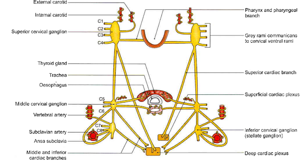
  • The cervical parts of the right and left sympathetic trunks 
  • are situated one on each side of the cervical part of the vertebral column, 
  • behind the carotid sheath
  • in front of the prevertebral fascia.

FORMATION
  • There are no white rami communicans in the neck 
  • this part of the trunk is formed by fibres which emerge from segments T1 to T4 of the spinal cord, and 
  • then ascend into the neck. 
  • Grey rami communicans (i.e. outgoing roots) are present.


RELATIONS
Anterior
a. Internal carotid artery
b. Common carotid artery
c. Carotid sheath
d. Inferior thyroid artery.

Posterior
a. Prevertebral fascia
b. Longus capitis and cervicis muscles
c. Transverse processes of the lower six cervical vertebrae.

Superior Cervical Ganglion
Size & Shape
This is the largest of the three ganglia. 
It is spindle shaped, and about 2.5 cm long.

Situation & Formation
It lies just below the skull, 
opposite the second and third cervical vertebrae, behind the carotid sheath and in front of the prevertebral fascia (longus capitis). 
It is formed by fusion of the upper 4 cervical ganglia.

Communication:
With cranial nerves IX, X and XII,
and with the external and recurrent laryngeal nerves

Branches
1. Grey rami communicans pass to the ventral rami of upper four cervical nerves.
2. The internal carotid nerve arises from the upper end of the ganglion and forms a plexus around the internal carotid artery. 
A part of this plexus supplies the dilator pupillae. 
Some of these fibres form the deep petrosal nerve for pterygopalatine ganglion; 
others give fibres along long ciliary nerve for the ciliary ganglion.
3. The external carotid branches form a plexus around the external carotid artery.
 Some of these fibres form the sympathetic roots of the otic and submandibular ganglia.
4. Pharyngeal branches take part in the formation of the pharyngeal plexus.
5. The left superior cervical cardiac branch goes to the superficial cardiac plexus while the right branch goes to the deep cardiac plexus.


Middle Cervical Ganglion
Size & Shape
This ganglion is very small. 
Situation
It lies in the lower part of the neck, in front of vertebra C6 just above the inferior thyroid artery, behind 
the carotid sheath.
Formation
It is formed by fusion of the fifth and sixth cervical ganglia connections. 
It is connected with the inferior cervical ganglion directly, and also through a loop that winds 
round the subclavian artery. This loop is called the ansa subclavia.


Branches
1. Grey rami communicans are given to the ventral rami of the 5th and 6th cervical nerves.
2. Thyroid branches accompany the inferior thyroid artery to the thyroid gland. They also supply the parathyroid glands.
3. Tracheal and oesophageal branches.
4. The middle cervical cardiac branch is the largest of the sympathetic cardiac branches. It goes to the deep cardiac plexus.


Inferior Cervical Ganglion
Size & Formation
It is formed by fusion of 7th and 8th cervical ganglia.
This is often fused with the first thoracic ganglion and is then known as the cervicothoracic ganglion or 
stellate ganglion because it is star-shaped.
It is situated between the transverse process of vertebra C7 and the neck of the first rib. 
It lies behind the vertebral artery, and in front of ramus of spinal nerve C8. 
A cervicothoracic ganglion extends in front of the neck of the first rib.

Branches
1. Grey rami communicans are given to the ventral rami of nerves C7 and C8.
2. Vertebral branches form a plexus around the vertebral artery.
3. Subclavian branches form a plexus around the subclavian artery. This plexus is joined by branches
from the ansa subclavia.
4. An inferior cervical cardiac branch goes to the deep cardiac plexus.


Clinical Anatomy:
The head and neck are supplied by sympathetic nerves arising from the upper four thoracic segments of the spinal cord. 
Most of these preganglionic fibres pass through the stellate ganglion to relay in the superior cervical ganglion.
Injury to cervical sympathetic trunk produces Horner's syndrome
It is characterized by:
a. Ptosis - drooping of the upper eyelid.
b. Miosis - constriction of the pupil.
c. Anhydrosis - loss of sweating on that side of the face.
d. Enophthalmos - retraction of the eyeball.
e. Loss of the ciliospinal reflex - pinching the skin on the nape of the neck does not produce dilatation of the pupil (which normally takes place).
Horner's syndrome can also be caused by a lesion within the central nervous system anywhere at or above the first thoracic segment of the spinal cord involving sympathetic fibres.


Watch the lecture on YouTube:


Thursday, July 23, 2020

Veins in the neck

SUBCLAVIAN VEIN
Course
It is a continuation of the axillary vein. 
It begins at the outer border of the first rib, and ends at the medial border of the scalenus anterior 
by joining the internal jugular vein to form the brachiocephalic vein.

It lies:
a. In front of the subclavian artery, the scalenus anterior and the right phrenic nerve
b. Behind the clavicle and the subclavius
c. Above the first rib and pleura.

Its tributaries are:
a. The external jugular vein
b. The dorsal scapular vein
c. The thoracic duct on the left side
d. The right lymphatic duct on the right side.

INTERNAL JUGULAR VEIN
Course
1. It is a direct continuation of the sigmoid sinus. 
It begins at the jugular foramen, and ends behind the sternal end of the clavicle by joining the subclavian vein to form the brachiocephalic vein.
2. The origin is marked by a dilation, the superior bulb which lies in the jugular fossa of he temporal bone,
beneath the floor of the middle ear cavity. 
The termination of the vein is marked by the inferior bulb which lies beneath the lesser supraclavicular fossa.


Relations
Superficial
1. Sternocleidomastoid
2. Posterior belly of digastric
3. Superior belly of omohyoid
4. Parotid gland
5.. Styloid process
6 The internal carotid artery, and the glossopharyngeal, vagus, accessory and hypoglossal cranial nerves
(at the base of skull).

Posterior
1. Transverse process of atlas
2. Cervical plexus
3. Scalenus anterior
4. First part of subclavian artery.

Medial
1. Internal carotid artery
2. Common carotid artery
3. Vagus nerve
Tributaries
1. Inferior petrosal sinus
2. Common facial vein
3. Lingual vein
4. Pharyngeal veins
5. Superior thyroid vein
6. Middle thyroid vein
The thoracic duct opens into the angle of union between the left internal jugular vein and the left
subclavian vein. 
The right lymphatic duct opens similarly on the right side.
In the middle of the neck, the internal jugular vein may communicate with the external jugular vein
through the oblique jugular vein which runs across the anterior border of the sternocleidomastoid.
Clinical Anatomy

Deep to the lesser supraclavicular fossa, the internal jugular vein is easily accessible for recording of venous pulse tracings. 

The vein can be cannulated by direct puncture in the interval between sternal and clavicular heads of sternocleidomastoid muscle.

In congestive cardiac failure or any other disease where venous pressure is raised, the internal jugular vein is markedly dilated and engorged.

BRACHIOCEPHALIC VEIN
1. The right brachiocephalic vein (2.5 cm long) is shorter than the left (6 cm long).
2. Each vein is formed behind the sternoclavicular joint,by the union of the internal jugular vein and the
subclavian vein.
3. The right vein runs vertically downwards. 
The left vein runs obliquely downwards and to the right behind the upper half of the manubrium sterni. 
The two brachiocephalic veins unite at the lower border of the right first costal cartilage to form the superior vena cava.
4. The tributaries correspond to the branches of the first part of the subclavian artery. 
These are as follows:
Right Brachiocephalic
a. Vertebral
b. Internal thoracic
c. Inferior thyroid
d. First posterior intercostal.

Left Brachiocephalic
a. Vertebral 
b. Internal thoracic
c. Inferior thyroid
d. First posterior intercostal.
e. Left superior intercostal.
f. Thymic and pericardial veins.

Watch the lectures on YouTube


Tuesday, July 21, 2020

Internal Carotid Artery

  • The internal carotid artery is one of the two terminal branches of the common carotid artery. 
  • It begins at the level of the upper border of the thyroid cartilage opposite the disc between the third and fourth cervical vertebrae, and 
  • ends inside the cranial cavity by supplying the brain. 
  • This is the principal artery of the brain and the eye. 
  • It also supplies the related bones and meninges.
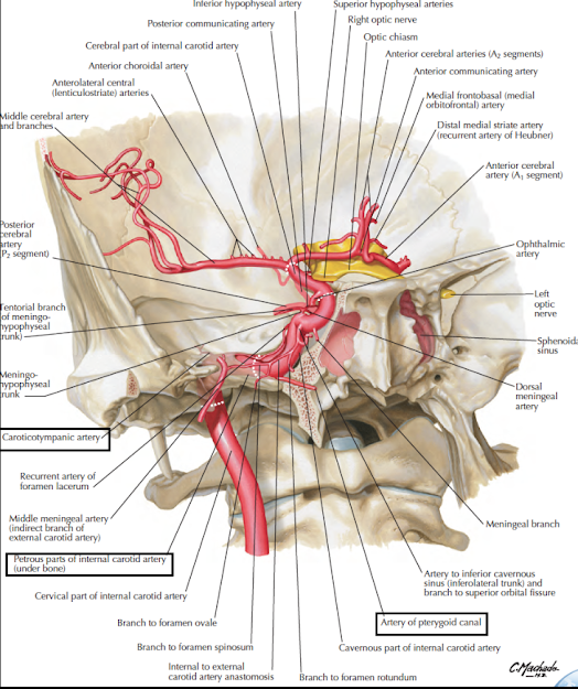
  • The course of the artery is divided into four parts:
a. Cervical part, in the neck
b. Petrous part, within the petrous temporal bone
c. Cavernous part, within the cavernous sinus
d. Cerebral part in relation to base of the brain.

Cervical Part
1. It ascends vertically in the neck from its origin to the base of the skull to reach the lower end of the carotid canal. 
This part is enclosed in the carotid sheath (with the internal jugular vein and the vagus).
2. No branches arise from the internal carotid artery in the neck
3. Its initial part usually shows a dilatation, the carotid sinus which acts as a baroreceptor 
4. The lower part of the artery (in the carotid triangle) is comparatively superficial. 
The upper part, above the posterior belly of the digastric, is deep to the parotid gland, the styloid apparatus, and many other structures.

Relations
Anterior or superficial
1. In the carotid triangle:
a. Anterior border of sternocleidomastoid
b. The external carotid artery is anteromedial to it.

2. Above the carotid triangle:
a. Posterior belly of the digastric
b. Stylohyoid
c. Stylopharyngeus
d. Styloid process
e. Parotid gland with structures within it.

Posterior
1. Superior cervical ganglion
2. Carotid sheath
3. The glossopharyngeal, vagus, accessory andhypoglossal nerves at the base of the skull.

Medial
1 Pharynx
2 The external carotid is anteromedial to it below the parotid.

Lateral
1. Internal jugular vein
2 Temporomandibular joint (at the base of the skull).

Petrous Part

1. In the carotid canal, the artery first runs upwards, and then turns forwards and medially at right angles.
It emerges at the apex of the petrous temporal bone, in the posterior wall of the foramen lacerum where
it turns upwards and medially.

2. Relations: The artery is surrounded by venous and sympathetic plexuses. 
It is related to the middle ear and the cochlea (posterosuperiorly); 
the auditory tube and tensor tympani (anterolaterally); and 
the trigeminal ganglion (superiorly).

3. Branches:
a. Caroticotympanic branches enter the middle ear, and anastomose with the anterior and posterior
tympanic arteries.
b. The pterygoid branch (small and inconstant) enters the pterygoid canal with the nerve of that 
canal and anastomoses with the greater palatine artery.

Watch the lectures o YouTube:


Monday, July 20, 2020

Subclavian Artery

Introduction
  • This is the principal artery which continues as axillary artery for the upper limb. 
  • It also supplies a considerable part of the neck and brain through its branches
Origin
  • On the right side, it is branch of the brachiocephalic artery. 
  • It arises posterior to the sternoclavicular joint.
  • On the left side, it is a branch of the arch of the aorta. 
  • It ascends and enters the neck posterior to the left sternoclavicular joint. 
  • Both arteries pursue a similar
  • course in the neck


Course
  • Each artery arches laterally from the sternoclavicular joint to the outer border of the first rib where it ends by becoming continuous with the axillary artery
  • The scalenus anterior muscle crosses the artery anteriorly and divides it into three parts. 
  • The first part is medial, the second part posterior, and the third part lateral to scalenus anterior.

Relations of the First Part

Anterior
Immediate relations from medial to lateral side are:
1. Common carotid artery
2. Vagus
3. Internal jugular vein
4. The sternothyroid and the sternohyoid muscles
5. Sternocleidomastoid.

Posterior 
1. Suprapleural membrane
2. Cervical pleura
3. Apex of lung.


Relations of the Second Part
Anterior
1. Scalenus anterior
2. Right phrenic nerve deep to the prevertebral fascia
3 Sternocleidomastoid.

Posterior (posteoinferior)
1. Suprapleural membrane
2. Cervical pleura
3. Apex of lung.

Superior
Upper and middle trunks of the brachial plexus
Relations of the third part
Anterior
1. Middle one-third of the clavicle
2. The posterior border of the sternocleidomastoid.

Posterior (Posteroinferior)
1. Scalenus medius
2. Lower trunk of brachial plexus
3 .Suprapleural membrane
4. Cervical pleura
5. Apex of lung.

Superior
Upper and middle trunks of brachial plexus.

lnferior
First rib
Branches
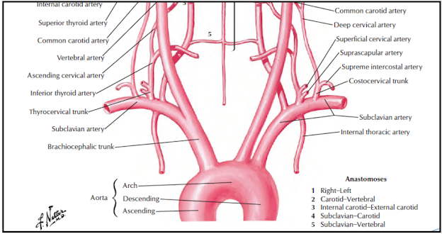
The subclavian artery gives off four branches. 
These are:
1. Vertebral artery.
2. Internal thoracic artery.
3. Thyrocervical trunk, which divides into threebranches:
    a. Inferior thyroid.
    b. Suprascapular.
    c. Transverse cervical arteries.
4 Costocervical trunk, which divides into two branches:
    a. Superior intercostal.
    b. Deep cervical arteries.
5 Dorsal scapular artery-occasionally.


Clinical Anatomy
  • The third part of the subclavian artery can be effectively compressed against the first rib after depressing the shoulder. 
  • The pressure is applied downwards, backwards, and medially in the angle between the sternocleidomastoid and the clavicie.
  • A cervical rib may compress the subclavian artery, diminishing the radial pulse.
  • The right subclavian artery may arise from the descending thoracic aorta. In that case, it passes posterior to  the oesophagus which may be compressed and the condition is known as (dysphagia lusoria).
  • An aneurysm may form in the third part of the subclavian artery. Its pressure on the brachial plexus causes pain, weakness/ and numbness in the upper limb.
  • Obstruction to the subclavian artery proximal to the origin of vertebral artery may lead to "stealing
  • of blood from the brain through the opposite vertebral artery. This may provide necessary blood to the affected side. The nervous symptoms incurred are called "subclavian steal syndrome"


Watch lectures on YouTube: