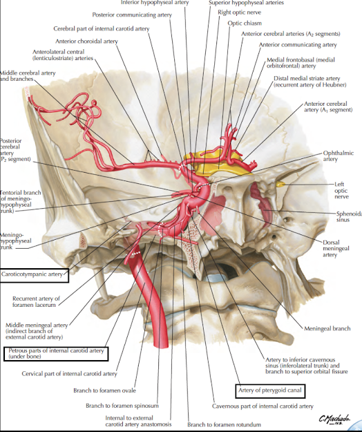
- The internal carotid artery is one of the two terminal branches of the common carotid artery.
- It begins at the level of the upper border of the thyroid cartilage opposite the disc between the third and fourth cervical vertebrae, and
- ends inside the cranial cavity by supplying the brain.
- This is the principal artery of the brain and the eye.
- It also supplies the related bones and meninges.
- The course of the artery is divided into four parts:
a. Cervical part, in the neck
b. Petrous part, within the petrous temporal bone
c. Cavernous part, within the cavernous sinus
d. Cerebral part in relation to base of the brain.
Cervical Part
1. It ascends vertically in the neck from its origin to the base of the skull to reach the lower end of the carotid canal.
This part is enclosed in the carotid sheath (with the internal jugular vein and the vagus).
2. No branches arise from the internal carotid artery in the neck
3. Its initial part usually shows a dilatation, the carotid sinus which acts as a baroreceptor
4. The lower part of the artery (in the carotid triangle) is comparatively superficial.
The upper part, above the posterior belly of the digastric, is deep to the parotid gland, the styloid apparatus, and many other structures.
Relations
Anterior or superficial
1. In the carotid triangle:
a. Anterior border of sternocleidomastoid
b. The external carotid artery is anteromedial to it.
2. Above the carotid triangle:
a. Posterior belly of the digastric
b. Stylohyoid
c. Stylopharyngeus
d. Styloid process
e. Parotid gland with structures within it.
Posterior
1. Superior cervical ganglion
2. Carotid sheath
3. The glossopharyngeal, vagus, accessory andhypoglossal nerves at the base of the skull.
Medial
1 Pharynx
2 The external carotid is anteromedial to it below the parotid.
Lateral
1. Internal jugular vein
2 Temporomandibular joint (at the base of the skull).
Petrous Part
1. In the carotid canal, the artery first runs upwards, and then turns forwards and medially at right angles.
It emerges at the apex of the petrous temporal bone, in the posterior wall of the foramen lacerum where
it turns upwards and medially.
2. Relations: The artery is surrounded by venous and sympathetic plexuses.
It is related to the middle ear and the cochlea (posterosuperiorly);
the auditory tube and tensor tympani (anterolaterally); and
the trigeminal ganglion (superiorly).
3. Branches:
a. Caroticotympanic branches enter the middle ear, and anastomose with the anterior and posterior
tympanic arteries.
b. The pterygoid branch (small and inconstant) enters the pterygoid canal with the nerve of that
canal and anastomoses with the greater palatine artery.
Watch the lectures o YouTube:


No comments:
Post a Comment