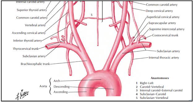
- This is the principal artery which continues as axillary artery for the upper limb.
- It also supplies a considerable part of the neck and brain through its branches
Origin
- On the right side, it is branch of the brachiocephalic artery.
- It arises posterior to the sternoclavicular joint.
- On the left side, it is a branch of the arch of the aorta.
- It ascends and enters the neck posterior to the left sternoclavicular joint.
- Both arteries pursue a similar
- course in the neck
Course
- Each artery arches laterally from the sternoclavicular joint to the outer border of the first rib where it ends by becoming continuous with the axillary artery
- The scalenus anterior muscle crosses the artery anteriorly and divides it into three parts.
- The first part is medial, the second part posterior, and the third part lateral to scalenus anterior.
Relations of the First Part
Anterior
Immediate relations from medial to lateral side are:
1. Common carotid artery
2. Vagus
3. Internal jugular vein
4. The sternothyroid and the sternohyoid muscles
5. Sternocleidomastoid.
Posterior
1. Suprapleural membrane
2. Cervical pleura
3. Apex of lung.
Relations of the Second Part
Anterior
1. Scalenus anterior
2. Right phrenic nerve deep to the prevertebral fascia
3 Sternocleidomastoid.
Posterior (posteoinferior)
1. Suprapleural membrane
2. Cervical pleura
3. Apex of lung.
Superior
Upper and middle trunks of the brachial plexus
Relations of the third part
Anterior
1. Middle one-third of the clavicle
2. The posterior border of the sternocleidomastoid.
Posterior (Posteroinferior)
1. Scalenus medius
2. Lower trunk of brachial plexus
3 .Suprapleural membrane
4. Cervical pleura
5. Apex of lung.
Superior
Upper and middle trunks of brachial plexus.
lnferior
First rib
Branches
The subclavian artery gives off four branches.
These are:
1. Vertebral artery.
2. Internal thoracic artery.
3. Thyrocervical trunk, which divides into threebranches:
a. Inferior thyroid.
b. Suprascapular.
c. Transverse cervical arteries.
4 Costocervical trunk, which divides into two branches:
a. Superior intercostal.
b. Deep cervical arteries.
5 Dorsal scapular artery-occasionally.
Clinical Anatomy
- The third part of the subclavian artery can be effectively compressed against the first rib after depressing the shoulder.
- The pressure is applied downwards, backwards, and medially in the angle between the sternocleidomastoid and the clavicie.
- A cervical rib may compress the subclavian artery, diminishing the radial pulse.
- The right subclavian artery may arise from the descending thoracic aorta. In that case, it passes posterior to the oesophagus which may be compressed and the condition is known as (dysphagia lusoria).
- An aneurysm may form in the third part of the subclavian artery. Its pressure on the brachial plexus causes pain, weakness/ and numbness in the upper limb.
- Obstruction to the subclavian artery proximal to the origin of vertebral artery may lead to "stealing
- of blood from the brain through the opposite vertebral artery. This may provide necessary blood to the affected side. The nervous symptoms incurred are called "subclavian steal syndrome"


No comments:
Post a Comment