- Largest mixed branch of trigeminal N
- Nerve of first pharyngeal arch
- Associated with Otic & submandibular ganglion
Begins in middle cranial fossa
It has a
- large sensory root
- arises from lateral part of trigeminal ganglion
- Leaves the cranial cavity through foramen ovale
- Passes deep to the trigeminal ganglion
- Passes through foramen ovale
- Joins with sensory root below the foramen forming the main trunk
- Lies in the infratemporal fossa, on the tensor veli palatini, deep to lateral pterygoid
- Then divides into anterior & posterior divisions
Branches from Main trunk
1. Meningeal N
- enters the cranial cavity through foramen spinosum
- Supplies the meninges of middle cranial fossa
2. N to medial pterygoid
- Supplies medial pterygoid
- Gives a motor root to otic ganglion without relay supplies tensor veli palatini & tensor tympani
Branches from anterior division
3 motor branches
- 1. Masseteric N
- 2. Deep temporal Ns
- 3. N to lateral pterygoid
1 sensory branch
- Buccal N
Branches from posterior division
Mostly sensory
1. Auriculotemporal N
- Arises by two root
- Encircles Middle meningeal A
- Auricular part – supplies skin of tragus, upper part of pinna, external acoustic meatus & tympanic membrane
- Temporal part – supplies temple, TMJ, carries secretomotor & sensory fibres to parotid gland
2. Lingual N
- One of the two terminal branches of posterior division
- Begins 1cm below the skull
- Runs b/n tensor veli palatini & lateral pterygoid.
- Then b/n lateral & medial pterygoid.
- 2cms below the skull, it is joined by chordatympani N
- Emerging at the lower border of lateral pterygoid
- Runs downwards & forwards b/n ramus of mandible & medial pterygoid
- Lies direct contact to mandible behind 3 molar tooth
- Runs over hyoglossus & genioglossus, deep to mylohyoid
- Winds around submandibular duct & divides into terminal branches
3. Inferior alveolar Nerve
- Larger terminal branch
- Runs vertically down lateral to the medial pterygoid & sphenomandibular ligament
- Enters mandibular foramen, runs in mandibular canal,
- Accompanied by inferior alveolar A
Branches
a. Mylohyoid branch
- only motor fibre from posterior division
- Pierces sphenomanibular ligament along with mylohyoid A
- Runs in mylohyoid groove
- Supplies mylohyoid & anterior belly of digastric muscle
- in the mandibular canal it gives branches to supply lower teeth & gums
- emerges from mental foramen, supplies skin of chin, skin & mucous membrane of lower lip.
- Its incisive branch supplies labial aspect of gums of canine & incisor teeth
Clinical Anatomy
- Motor part of mandibular N is tested by asking the patient to clench teeth. If one side masseter paralyzed, jaw deviates to paralyzed side while opening the mouth by the action of lateral pterygoid of opposite side.
- Referred pain – in case of cancer of tongue, pain radiates to the ear & temporal fossa along the distribution of auriculotemporal N, lingual N
- Lingual N is divided to relieve intractable pain. Done where the nerve lies in contact with mandible below & behind the 3 molar tooth.
Trigeminal neuralgia
- Intractable pain along distribution of mandibular N
- In this sensory root may be divided behind the ganglion
- During division ophthalmic fibres lie in the superomedial part of root are spared to preserve the corneal reflex
- In extraction of wisdom tooth lingual N should be taken care. Its injury results sensory loss in anterior 2/3 of tongue
- A lesion at foramen ovale leads to paraesthesia along mandible, tongue, temporal region, paralysis of muscles of mastication, loss of jaw jerk
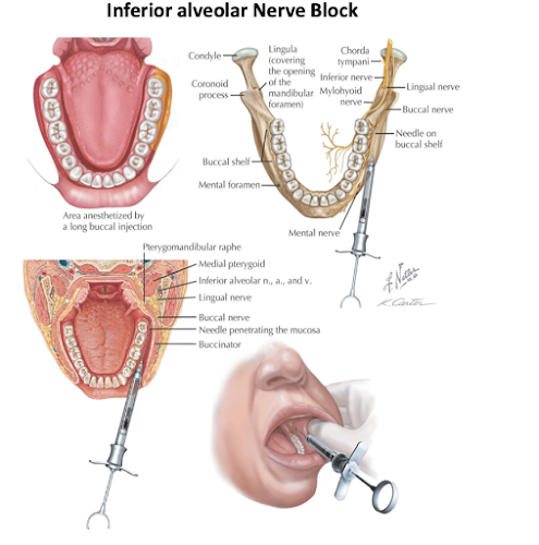
- In extraction of tooth, inferior alveolar N needs to be anaesthetized. The drug is given before the N enters the mandibular canal.
- Inferior alveolar N can be damaged in the fracture of mandible
- During extraction of 3 molar tooth, buccal N may be involved resulting in temporary numbness of cheek
Auriculo-temporal nerve - Course | Distribution | Clinical Anatomy
Lingual Nerve | Course | Relations | Distribution | Clinical Anatomy
Lingual Nerve | Course | Relations | Distribution | Clinical Anatomy


Very informative notes.
ReplyDeleteThank U
Delete