Features
The lateral wall of the nose is irregular owing to the presence of three shelf-like bony projections called conchae.
The conchae increase the surface area of the nose for effective air-conditioning of the inspired air
The lateral wall separates the nose:
a. From the orbit above, with the ethmoidal air sinuses intervening.
b. From the maxillary sinus below.
c. From the lacrimal groove and nasolacrimal canal in front.
The lateral wall can be subdivided into three parts.
a. A small depressed area in the anterior part is called the vestibule.
It is lined by modified skin containing short, stiff, curved hairs called vibrissae.
b. The middle part is known as the atrium of the middle meatus.
c. The posterior part contains the conchae.
Spaces separating the conchae are called meatuses

The skeleton of the lateral wall is partly bony, partly cartilaginous, and partly made up only of soft tissues.
The bony part is formed from before backwards by the following bones:
a. Nasal.
b. Frontal process of maxilla.
c. Lacrimal.
d. Labyrinth of ethmoid with with superior and middle conchae.
e. Inferior nasal concha made up of spongy bone only
f . Perpendicular plate of palatine bone together with its orbital and sphenoidal processes.
g. Medial pterygoid plate.
The cartilaginous part is formed by:
a. The superior nasal cartilage.
b. The inferior nasal cartilage.
c. 3 or 4 small cartilages of the ala
The cuticular lower part is formed by fibrofatty tissue covered with skin

Chonchae
Features
The nasal conchae are curved bony projections directed downwards and medially.
The following three conchae are usually found:
1. The inferior concha is an independent bone.
2. The middle concha is a projection from the medial surface of ethmoidal labyrinth
3. The superior concha is also a projection from the medial surface of the ethmoidal labyrinth.
This is the smallest concha situated just above the posterior part of the middle concha

Meatuses
- The meatuses of the nose are passages beneath the overhanging conchae.
- Each meatus communicates freely with the nasal cavity proper.
1. The inferior meatus lies underneath the inferior concha, and is the largest of the three meatuses.
• The nasolacrimal duct opens into it at the junction of its
anterior one-third and posterior two-thirds.
• The opening is guarded by the lacrimal fold, or Hasner's valve.
2 The middle meatus lies underneath the middle concha.
It presents the following features:
a. The ethmoidal bulla, is a rounded elevation produced by the underlying middle ethmoidal sinuses which open at upper margin of bulla.
b. The hiatus semilunaris, is a deep semicircular sulcus below the bulla.
c. The infundibulum is a short passage at the anterior end of the hiatus.
d. The opening of frontal air sinus is seen in the anterior part of hiatus semilunaris
e. The opening of the anterior ethmoidal air sinus is present behind the opening of frontal air sinus.
f . The opening of maxillary air sinus is located in posterior part of the hiatus semilunaris.
It is often represented by two openings.

3. The superior meatus lies below the superior concha.
This is the shortest and shallowest of the three meatuses.
It receives the openings of the posterior ethmoidal air sinuses.
The sphenoethmoidal recess is a triangular fossa just
It receives lhe opening of the sphenoidal air sinus
The atrium of the middle meatus is a shallow depression just in front of the middle meatus and above the vestibule of the nose.
It is limited above by a faint ridge of mucous membrane, the agger nasi, which runs forwards and downwards from the upper end of the anterior border of the middle concha

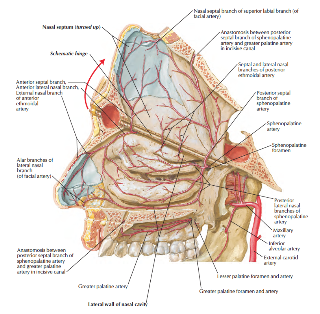
Arterial Supply
1. The anterosuperior quadrant is supplied by the anterior ethmoidal artery assisted by the posterior ethmoidal artery.
2. The anteroinferior quadrant, is supplied by branches from the facial artery.
3. The posterosuperior quadrant, is supplied by few branches of the sphenopalatine artery
4 The posteroinferior quadrant is supplied by branches from greater palatine artery which pierce the perpendicular plate of palatine bone and passes up through the incisive fossa.

Venous Drainage
The veins form a plexus which drains
• anteriorly into the facial vein;
• posteriorly, into the pharyngeal plexus of veins; and
• from the middle part, to the pterygoid plexus of veins.

Nerve Supply
General sensory nerves derived from the branches of trigeminal nerve are distributed to whole of the lateral wall:
a. Anterosuperior quadrant is supplied by the anterior ethmoidal nerve branch of ophthalmic nerve b. Anteroinferior quadrant is supplied by the anterior superior alveolar nerve, branch of infraorbital, continuation of maxillary nerve.
c. Posterosuperior quadrant is supplied by the lateral posterior superior nasal branches from the pterygopalatine ganglion.
d. Posteroinferior quadrant is supplied by the anterior palatine branch from the pterygopalatine ganglion.
Special sensory nerves or olfactory nerves are distributed to the upper part of the lateral wall just below the cribriform plate of the ethmoid up to the superior concha.

Lymphatic Drainage
Lymphatics from the anterior half of the lateral wall pass to the submandibular nodes, and
from the posterior half, to the retropharyngeal and upper deep cervical nodes.
Clinical Anatomy
Hypertrophy of the mucosa over the inferior nasal concha is a common feature of allergic rhinitis, which is characterized by sneezing, nasal blockage and excessive watery discharge from the nose
Watch Lectures on YouTube:
Lateral Wall of Nasal cavity | Formation | Conchae | Meatuses | Air sinuses Opening | Blood Supply
No comments:
Post a Comment