Features
The nasal septum is median osseocartilaginous partition between the two halves of the nasal cavity.
On each side, it is covered by mucous membrane and forms the medial wall of both nasal cavities.
The bony part is formed almost entirely by:
a. The vomer, and
b. The perpendicular plate of ethmoid.
However, its margins receive contributions from the nasal spine of the frontal bone, the rostrum of the sphenoid, and the nasal crests of the nasal, palatine and maxillary bones.
The cartilaginous part is formed by:
a. The septal cartilage, and
b. The septal processes of the inferior nasal cartilages
The cuticular part or lower end is formed by fibrofatty tissue covered by skin.
The lower margin of the septum is called the columella.
The nasal septum is rarely strictly median.
Its central part is usually deflected to one or the other side.
The deflection is produced by overgrowth of one or more of the constituent parts.
The septum has:
a. Four borders - superior, inferior, anterior and posterior.
b. Two surfaces - right and left.

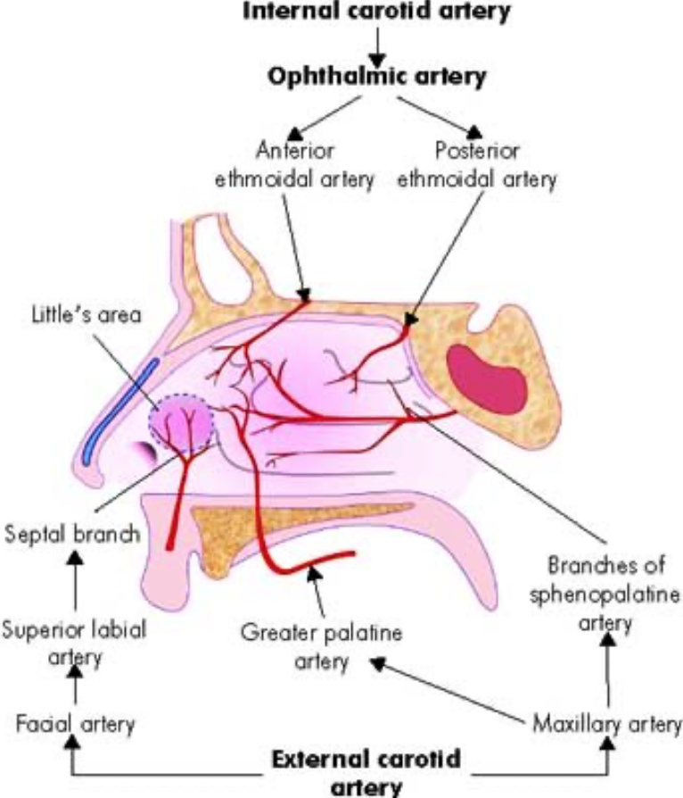
Arterial Supply
Anterosuperior part is supplied by the anterior and posterior ethmoidal arteries.
Anteroinferior part by the superior labial branch of facial artery.
Posterosuperior part is supplied by the sphenopalatine artery. It is the main artery.
Posteroinferior part by branches of greater palatine artery.
The anteroinferior part or vestibule of the septum contains anastomoses between the septal ramus of the
superior labial branch of the facial artery, branch of sphenopalatine artery, greater palatine and of anterior
ethmoidal artery.
These form a large capillary network called the Kiesselbach's plexus.
This is a common site of bleeding from the nose or epistaxis, and is known as Little's area.

Venous Drainage
The veins form a plexus which is more marked in the lower part of septum or Little's area.
The plexus drains anteriorly into the facial vein, posteriorly through the sphenopalatine vein to pterygoid venous plexus.

Nerve Supply
General sensory nerves, arising from trigeminal nerve, are distributed to whole of the septum
a. The anterosuperior part of the septum is supplied by the internal nasal branches of the anterior ethmoidal nerve.
b. Its anteroinferior part is supplied by anterior superior alveolar nerve.
c. The posterosuperior part is supplied by the medial posterior superior nasal branches of the pterygopalatine ganglion
d. The posteroinferior part is supplied by the nasopalatine branch of the pterygopalatine ganglion. lt is the main nerve.
Special sensory nerves or olfactory nerves are confined to the upper part or olfactory area.

Lymphatic Drainage
Anterior half to the submandibular nodes.
Posterior half to the retropharyngeal and deep cervical nodes.
Clinical Anatomy
Sphenopalatine artery is the artery of epistaxis.
Little's area on the septum is a common site of bleeding from the nose or epistaxis.
Pathological deviation of the nasal septum is often responsible for repeated attacks of common cold, allergic rhinitis, sinusitis, etc.
It requires surgical correction
Watch lectures on YouTube:
Medial Wall of Nasal Cavity /Nasal Septum | Bones & Cartilages | Blood supply | Nerve Supply
No comments:
Post a Comment