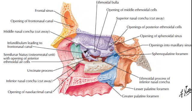
Features
- Paranasal sinuses are air filled spaces present within some bones around the nasal cavities.
- The sinuses, are frontal, maxillary, sphenoidal and ethmoidal.
- All of them open into the nasal cavity through its lateral wall
- The function of the sinuses is to make the skull lighter and add resonance to the voice.
- In infections of the sinuses or sinusitis, the voice is altered.
- The sinuses are rudimentary, or even absent at birth.
- They enlarge rapidly during the ages of 6 to 7 years, i.e. time of eruption of permanent teeth and then after puberty.
- From birth to adult life, the growth of the sinuses is due to enlargement of the bones;
- in old age it is due to resorption of the surrounding cancellous bone.
Frontal Sinus
1. The frontal sinus lies in the frontal bone deep to the superciliary arch.
It extends upwards above the medial end of the eyebrow, and backwards into the medial part of the roof of the orbit.
2. It opens into the middle meatus of nose at the anterior end of the hiatus semilunaris either through the infundibulum or through the frontonasal duct
3. The right and left sinuses are usually unequal in
size; and
rarely one or both may be absent.
Their average height, width and anteroposterior depth are each about 2.5 cm.
The sinuses are better developed in males than in females.
4. They are rudimentary or absent at birth.
They are well developed between 7 and 8 years of age, but reach full size only after puberty.
5. Arterial supply: Supraorbital artery.
6. Venous drainage: Into the supraorbital and superior ophthalmic veins.
7. Lymphatic drainage: To submandibular nodes.
8. Nerve supply: Supraorbital nerve.
Maxillary Sinus
1. The maxillary sinus lies in the body of the maxilla and is the largest of all the paranasal sinuses.
It is pyramidal in shape, with its base directed medially towards the lateral wall of the nose, and the apex directed laterally in the zygomatic process of the maxilla.
2. It opens into the middle meatus of the nose in the lower part of the hiatus semilunaris.
The opening is nearer the roof.
3. In an isolated maxilla, the opening or hiatus of the maxillary sinus is large.
However, in the intact skull the size of opening is reduced
to 3 or 4 mm as it is overlapped by the following:
a. From above, by the uncinate process of the ethmoid, and the descending part of lacrimal bone.
b. From below, by the inferior nasal concha.
c. From behind, by the perpendicular plate of the palatine bone.
It is further reduced in size by the thick mucosa of nose.
4. The size of sinus is variable.
Average measurements are: height - 3.5 cm, width - 2.5
cm and anteroposterior depth -3.5 cm.
5. Its roof is formed by the floor of orbit, and is traversed by the infraorbital nerve.
The floor is formed by the alveolar process of maxilla, and
lies about 1 cm below the level of floor of the nose. The level corresponds to the level of lower border of the ala of nose.
The floor is marked by several conical elevations
produced by the roots of upper molar and premolar
teeth.
The roots may even penetrate the bony floor to lie beneath the mucous lining.
The canine tooth may project into the anterolateral wall.
The maxillary sinus is the first paranasal sinus to
Arterial supply: Facial, infraorbital and greater palatine arteries.
Venous drainage into the facial vein and the pterygoid plexus of veins.
Lymphatic drainage into the submandibular nodes.
Nerve supply; Posterior superior alveolar nerves from maxillary and anterior and middle superior alveolar nerves from infraorbital.
Sphenoidal Sinus:
The right and left sphenoidal sinuses lie within the body of sphenoid bone
They are separated by a septum
The two sinuses are usually unequal in size Each sinus opens into sphenoethmoidal recess of corresponding half of the nasal cavity. Each sinus is related superiorly to the optic chiasma and the hypophysis cerebri; and laterally to the internal carotid artery and the cavernous sinus
Arterial supply: Posterior ethmoidal and internal carotid arteries.
Venous drainage: Into pterygoid venous plexus and cavernous sinus.
Lymphatic drainage: To the retropharyngeal nodes.
Nerve supply: Posterior ethmoidal nerve and orbital branches of pterygopalatine ganglion.
Ethmoidol Sinuses
• Ethmoidal sinuses are numerous small intercommunicating
spaces which lie within the labyrinth of the ethmoid bone.
- They are completed from above by the orbital plate of the
frontal bone, - from behind by the sphenoidal conchae and the orbital
plate of frontal bone and - anteriorly by the lacrimal bone.
- The sinuses are divided anterior, middle and posterior
groups. - The anterior ethmoidal sinus is made up of 1 to 11 air
cells, - opens into the anterior part of the hiatus semilunaris of the
nose. - It is supplied by the anterior ethmoidal nerve and vessels.
- Its lymphatics drain into the submandibular nodes.
- The middle ethmoidal sinus consisting of 1 to 7 air cells
- open into the middle meatus of the nose. It is
- supplied by the anterior ethmoidal nerve and vessels and
the orbital branches of the pterygopalatine ganglion. - Lymphatics drain into the submandibular nodes.
- The posterior ethmoidal sinus consisting of 1 to 7 air cells
- open into the superior meatus of the nose.
- It is supplied by the posterior ethmoidal nerve and vessels
and the orbital branches of the pterygopalatine ganglion. - Lymphatics drain into the retropharyngeal nodes.
Clinical Anatomy
- Infection of a sinus is known as sinusitis. It causes headache and persistent, thick, purulent discharge from the nose. Diagnosis is assisted by transillumination and radiography. A diseased sinus is opaque.
- The maxillary sinus is most commonly involved. It may be infected from the nose or from a caries tooth. Drainage of the sinus is difficult because its ostium lies at a higher level than its floor.
- Hence, the sinus is drained surgically by making an artificial opening near the floor in one of the following two ways:
a. Antrum puncture can be done by breaking the lateral wall of the inferior meatus and pushing in fluid and letting it drain through the natural orifice with head in dependent position
b. An opening can be made at the canine fossa through the vestibule of the mouth, deep to the upper lip (Caldwell-Luc operation).
• Carcinoma of the maxillary sinus arises from the mucosal lining. Symptoms depend on the direction of growth.
a. Invasion of the orbit causes proptosis and diplopia. If the infraorbital nerve is involved, there is facial pain and anaesthesia of the skin over the maxilla.
b. Invasion of the floor may produce a bulging and even ulceration of the Palate.
c. Forward growth obliterates the canine fossa and produces a swelling of the face.
d. Backward growth may involve the palatine nerves and produce severe pain referred to the upper teeth.
e. Growth in a medial direction produces nasal obstruction, epistaxis and epiphora.
f. Growth in a lateral direction produces a swelling on the face and a palpable mass in the labiogingival groove.
• Frontal sinusitis and ethmoiditis can cause oedema of the lids secondary to infection of the sinuses.
• Pain from ethmoid air sinus may be referred to forehead, as both are supplied by ophthalmic division of trigeminal nerve.
• Pain of maxillary sinusitis may be referred to upper teeth and infraorbital skin as all these are supplied by the maxillary nerve.


No comments:
Post a Comment